CBCT Imaging for Root Canal Treatment: How 3D Scans Reveal Hidden Problems

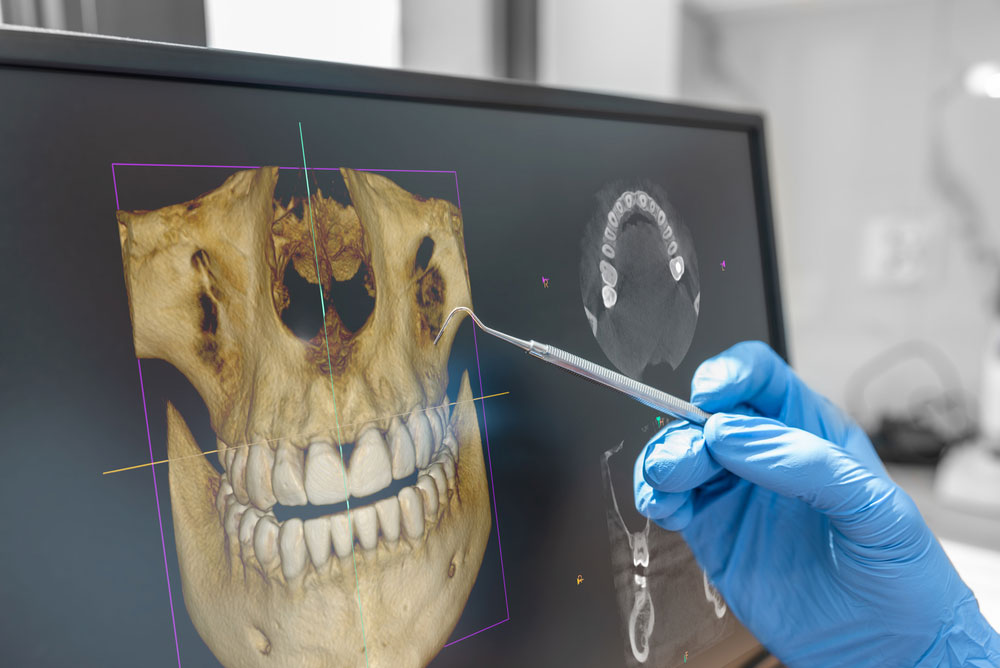
A standard dental X-ray can show a great deal, but some tooth problems hide behind overlapping roots, dense bone, or complex internal anatomy. In selected cases, CBCT imaging for root canal treatment gives an endodontist a three-dimensional view that can make diagnosis and planning more precise. Lake Houston Endodontics uses advanced dental technologies in Humble, TX to investigate difficult symptoms and plan specialized care.

A CBCT scan is not needed for every toothache or every root canal. Instead, it is used when the additional information may change the diagnosis, reveal anatomy that cannot be seen clearly on two-dimensional images, or help the endodontist choose between treatment options. Understanding what the scan does can help patients feel more confident when CBCT imaging for root canal treatment is recommended.

What Is a Dental CBCT Scan?

CBCT stands for cone beam computed tomography. The scanner moves around the patient’s head and captures many small images from different angles. Computer software then combines those images into a three-dimensional view of the teeth, roots, jawbone, and nearby structures.

The resulting data can be viewed in thin slices from several directions. An endodontist may rotate the image, enlarge a particular root, or examine the area from the side, front, and top. This flexibility is valuable because root canal systems are three-dimensional, even though traditional dental X-rays flatten the area into a two-dimensional picture.

CBCT imaging for root canal treatment is focused on diagnosis and planning. It does not replace a clinical examination, pulp testing, bite testing, or standard digital X-rays. Instead, it adds another layer of information when the basic findings leave an important question unanswered.

How CBCT Differs From a Standard Dental X-Ray

A standard dental X-ray is quick, useful, and appropriate for many endodontic evaluations. It can show decay, large restorations, bone changes, previous root canal fillings, and the general shape of roots. However, structures can overlap. A root positioned behind another root may be difficult to separate, and a small area of bone loss may not be visible from the angle used.

CBCT imaging for root canal treatment reduces the problem of overlap by displaying the area in three dimensions. For example, the endodontist may be able to see whether a dark area is associated with one root or another. The scan may also show that a root curves in a direction that was hidden on the standard image.

Still, more imaging is not always better. A standard X-ray may already provide everything needed for a straightforward case. The endodontist chooses CBCT when the expected diagnostic value justifies the scan.

Finding Extra or Unusual Root Canals

Teeth do not all follow a simple blueprint. A molar may have an extra canal, a canal may divide and reconnect, or roots may curve sharply. These variations can be difficult to identify on a flat image. If infected tissue remains in an untreated canal, symptoms or disease may continue after treatment.

CBCT imaging for root canal treatment can help map unusual anatomy before the procedure begins. This gives the endodontist a clearer idea of where to look and how the canals may travel through the root. Magnification and illumination are then used during treatment to locate and clean the actual openings.

The scan does not perform the treatment, and it cannot guarantee that every microscopic branch will be visible. However, a three-dimensional roadmap can make a complex case more understandable and support a more efficient plan.

Evaluating a Tooth That Still Hurts After a Root Canal

A previously treated tooth may become painful months or years later. Causes can include a missed canal, leakage from a restoration, new decay, a fracture, or persistent inflammation around a root. Because root filling materials and crowns can make standard images harder to interpret, additional views may be useful.

CBCT imaging for root canal treatment can show the location and size of bone changes around individual roots. It may also reveal untreated anatomy or a problem that affects only one side of a tooth. These details help the endodontist determine whether endodontic retreatment, surgery, monitoring, or another option deserves consideration.

Not every source of pain appears on a scan. Bite problems, muscle pain, early cracks, and some inflammatory conditions may require clinical testing and careful history. Therefore, imaging is interpreted as part of the whole examination rather than treated as the only answer.

Planning Endodontic Surgery

When inflammation remains near a root tip after nonsurgical treatment, an apicoectomy or another form of endodontic surgery may be considered. Before surgery, the endodontist needs to understand the root’s position, nearby bone, and relationship to surrounding anatomy.

CBCT imaging for root canal treatment and surgery can help identify which root is involved and where the problem is located. It may also show the thickness of bone over the root or the proximity of nearby structures. This information supports a more targeted surgical approach.

Patients can learn more about endodontic surgery in Humble, TX when nonsurgical treatment alone cannot address the problem. The recommendation depends on the tooth, restorability, and expected long-term outlook.

Can CBCT Find a Cracked Tooth?

Cracks are among the most challenging dental problems to diagnose. A large fracture or the bone pattern associated with a root fracture may be visible on CBCT. However, very fine cracks can still be too small to appear clearly. Restorations and root filling materials may also create visual artifacts that make interpretation harder.

For this reason, CBCT imaging for root canal treatment is only one part of a cracked tooth evaluation. Bite testing, magnification, transillumination, gum measurements, and symptom patterns may provide important clues. Pain when releasing a bite, isolated deep gum probing, or a recurring gum bump can influence the diagnosis even if the crack itself is not visible.

A scan can still be valuable because it may rule in or rule out other causes. For example, it may show a missed canal or localized bone loss that changes the treatment discussion.

Benefits of CBCT Imaging for Root Canal Treatment

  • Three-dimensional detail: The scan allows roots and surrounding bone to be viewed from multiple directions.
  • Improved anatomy mapping: CBCT imaging for root canal treatment may reveal extra canals, unusual curves, or root variations.
  • More focused retreatment planning: The endodontist can identify which root or region may be associated with persistent disease.
  • Better surgical preparation: A scan can clarify the location of a root tip and nearby structures before endodontic surgery.
  • Support for difficult diagnosis: CBCT may provide useful information when symptoms continue but standard images are inconclusive.
  • Clearer patient communication: Three-dimensional views can help patients understand why a treatment option is being recommended.

What the Scan Experience Is Like

CBCT imaging is generally quick. You may stand or sit while the machine is positioned around your head. The team asks you to remain still, and the scanner rotates around the area. Nothing is placed deep inside the mouth, although positioning supports may be used to help you stay steady.

The scan itself does not hurt. After the image is captured, the endodontist reviews the data on a computer. In many cases, the most relevant views can be shown during the consultation so you can see the roots and surrounding bone.

Metal jewelry, removable appliances, or other objects near the head may interfere with the image. Follow the team’s instructions about removing earrings, glasses, or removable dental items before the scan.

How Imaging Is Selected Thoughtfully

CBCT imaging for root canal treatment is ordered when the expected benefit is meaningful. The field of view can often be limited to the region being evaluated, rather than scanning a larger area than necessary. The dental team also considers whether previous imaging is available and whether a standard X-ray can answer the question.

Patients should tell the office if they are pregnant or may be pregnant before imaging. They should also mention recent scans so the endodontist can determine whether those records may be useful. If another dental office has already taken a CBCT scan, ask whether the file can be transferred for review.

It is reasonable to ask why the scan is recommended. A clear answer might include locating an extra canal, evaluating a root fracture, measuring a bone lesion, or planning surgery. The reason should be connected to a specific diagnostic or treatment decision.

How CBCT Works With Microscopes and Digital X-Rays

Endodontic technology is most effective when tools complement one another. Digital X-rays provide efficient two-dimensional views. CBCT offers three-dimensional context. Meanwhile, a dental microscope gives high magnification and illumination inside the tooth during treatment.

For example, CBCT imaging for root canal treatment may suggest that a narrow extra canal is present. The microscope then helps the endodontist search for the canal opening. Ultrasonic instruments may be used to remove small amounts of calcified material while preserving surrounding tooth structure.

No technology replaces training and judgment. The endodontist must decide which finding matters, whether it matches the symptoms, and whether treatment can improve the tooth’s outlook.

Frequently Asked Questions About CBCT Imaging for Root Canal Treatment

Does every root canal need a CBCT scan?

No. Many root canals can be diagnosed and treated with an examination and standard digital X-rays. CBCT is used when three-dimensional information may improve diagnosis or planning.

Can a CBCT scan show infection?

It can show changes in the bone that may be associated with inflammation or infection. The endodontist combines those findings with symptoms, testing, and the condition of the tooth.

Will CBCT always show a crack?

No. Some cracks are too fine to be seen directly, and metal or filling materials can affect the image. Clinical tests remain important in a cracked tooth evaluation.

Is the scan uncomfortable?

The scan is generally quick and painless. The main requirement is remaining still while the machine rotates around the head.

Can an old CBCT scan be used?

Possibly. The value depends on when it was taken, the area included, image quality, and whether the tooth has changed since then. The endodontist can review the file and decide whether new imaging is needed.

See the Full Picture Before Treatment

CBCT imaging for root canal treatment can reveal details that are hidden by overlapping anatomy on a standard X-ray. When used selectively, it helps an endodontist investigate complex pain, plan retreatment, evaluate surgery, and explain findings more clearly. The scan is most useful when it answers a specific question and is interpreted alongside a complete clinical examination.

For advanced endodontic diagnosis in Humble, TX, contact Lake Houston Endodontics. Call Us Today at (832) 777-6056 or contact the practice online to schedule an appointment and learn whether CBCT imaging for root canal treatment may be appropriate for your tooth.


Root Canal Treatment During Pregnancy in Humble, TX: Safety, Timing, and Comfort

A sudden toothache can be stressful at any time, but it may feel especially complicated when you are expecting a baby. Many patients wonder whether they should delay care, avoid dental X-rays, or try to manage the pain until after delivery. In reality, untreated infection can create its own concerns, so a timely evaluation is important. Lake Houston Endodontics provides root canal treatment in Humble, TX for patients who need focused care to relieve pain and preserve a natural tooth.

Root canal treatment during pregnancy is planned with your comfort, stage of pregnancy, medical history, and dental condition in mind. Your endodontist can also coordinate with your obstetric provider when additional guidance is helpful. The goal is not to rush into treatment or postpone necessary care without a reason. Instead, the goal is to make a thoughtful decision based on the health of the tooth and the needs of the patient.

Why Tooth Pain Should Be Evaluated During Pregnancy

Pregnancy brings many physical changes, and dental symptoms can sometimes be easy to dismiss as one more temporary discomfort. However, a tooth that throbs, keeps you awake, reacts strongly to heat, or causes swelling may have inflammation or infection inside it. These problems usually do not resolve simply because a patient avoids chewing on the area.

When the pulp inside a tooth becomes severely inflamed or infected, the source is enclosed within the hard structure of the tooth. Therefore, brushing more often or taking medication may reduce symptoms for a short time, but those steps do not remove damaged tissue from the root canal system. An examination can determine whether the tooth needs monitoring, restorative care, or root canal treatment during pregnancy.

Prompt evaluation is also useful because dental pain can affect eating, sleeping, hydration, and daily comfort. In addition, swelling can worsen quickly. Contact a dental professional promptly if you notice facial swelling, drainage, fever, a gum bump, a bad taste, or difficulty swallowing. Severe or spreading symptoms need urgent attention.

Is Root Canal Treatment During Pregnancy Possible?

Yes, root canal treatment during pregnancy can be performed when it is needed. Dental care is individualized, so the timing and details depend on the patient’s health, the condition of the tooth, and the urgency of the symptoms. A painful or infected tooth may require care during any trimester, especially when delaying treatment could allow the problem to become more difficult to manage.

For nonurgent procedures, many patients find the middle portion of pregnancy more comfortable because early nausea may have improved and lying back may still be manageable. However, a dental emergency does not always follow an ideal calendar. If a tooth is causing severe pain or infection, the dental team can discuss how to provide appropriate care rather than asking the patient to endure symptoms for months.

Before the appointment, share your due date, pregnancy history, allergies, current medications, and any recommendations from your obstetric provider. Also mention complications such as high blood pressure, bleeding concerns, or a pregnancy that requires special monitoring. Complete information helps the endodontist plan root canal treatment during pregnancy more carefully.

What About Dental X-Rays?

Endodontic diagnosis depends on understanding the roots, surrounding bone, and location of infection. Digital dental X-rays provide focused images that help the endodontist determine which tooth is responsible and how its canals are shaped. Without appropriate imaging, it may be difficult to diagnose the problem or treat the tooth precisely.

Modern dental imaging is directed at a small area. The dental team also follows safety procedures and uses imaging only when it contributes meaningful information. If you are pregnant or think you may be pregnant, tell the office before any images are taken. The team can explain why a particular image is recommended and how it will influence care.

In selected complex cases, additional imaging may be discussed. However, advanced scans are not automatically needed for every root canal treatment during pregnancy. The endodontist weighs the value of the information against the patient’s circumstances and chooses the least amount of imaging that can answer the clinical question.

Local Anesthetic and Comfort During Treatment

A root canal should not be performed without appropriate numbing. Local anesthetic helps keep the treatment area comfortable while the inflamed or infected tissue is removed. Your endodontist will review your medical history and choose an approach that fits your needs.

Some pregnant patients worry that they should avoid all medication, including local anesthetic. However, trying to complete treatment without adequate numbing can create unnecessary stress and make the procedure harder to tolerate. The safer approach is to discuss concerns openly and allow the dental and medical teams to guide medication decisions.

Positioning also matters, especially later in pregnancy. A patient may be more comfortable with the chair adjusted slightly, a small cushion for support, or brief breaks during a longer appointment. Tell the team if you feel lightheaded, short of breath, nauseated, or uncomfortable. Root canal treatment during pregnancy should be adapted to the patient rather than expecting the patient to remain silent through discomfort.

Medication Planning Before and After Care

Medication choices during pregnancy require individual review. Do not start, stop, or change a prescription based only on general information online. Instead, provide the endodontic team with a complete list of medications and supplements. The office may coordinate with your obstetric provider when a pain reliever, antibiotic, or sedative requires additional discussion.

Antibiotics are not automatically prescribed for every root canal. When the infection is contained within the tooth and there are no signs of spread, treatment of the source may be the main priority. In other situations, swelling, fever, or other findings may affect the plan. The endodontist will decide based on the examination rather than prescribing medication simply because a tooth hurts.

Dental anxiety should also be discussed before the appointment. Lake Houston Endodontics offers sedation dentistry, but pregnancy changes how sedation options are considered. The safest plan may involve local anesthetic, reassurance, shorter appointments, and nonmedication comfort techniques. Any sedation decision should be made only after a careful medical review.

What Happens During Root Canal Treatment During Pregnancy?

The procedure itself follows the same tooth-saving principles used for other patients. First, the endodontist examines the tooth and reviews the images. After the tooth is numb, a protective dental dam is placed around it. This small sheet isolates the tooth, helps keep the treatment area clean, and prevents materials from entering the mouth.

Next, a small opening is made in the tooth. Fine instruments are used to remove inflamed or infected pulp, clean the canals, and shape the internal spaces. The canals are disinfected and sealed with a biocompatible filling material. Depending on the tooth and diagnosis, treatment may be completed in one visit or divided between appointments.

A temporary filling is often placed before the patient returns to the general dentist for a permanent restoration. That final restoration is important because root canal treatment addresses the inside of the tooth, while a crown or filling protects the remaining tooth structure from chewing forces and leakage.

Benefits of Root Canal Treatment During Pregnancy

  • Relief from the source of pain: Treatment removes inflamed or infected tissue rather than only masking discomfort.
  • Protection of the natural tooth: Root canal treatment during pregnancy may allow a tooth to remain in place instead of being extracted.
  • Improved eating and sleeping: Addressing severe tooth pain can make daily routines more comfortable.
  • Reduced risk of worsening infection: Timely care can help prevent a localized problem from becoming a larger emergency.
  • Personalized medication planning: The dental team can select an approach based on pregnancy, medical history, and provider guidance.
  • Focused specialty care: An endodontist is trained to diagnose complex tooth pain and treat the root canal system precisely.

How to Prepare for the Appointment

Start by giving the office accurate medical information. Bring the name and contact information of your obstetric provider, a list of medications, your due date, and any special instructions you have received. If your pregnancy is considered high risk, let the office know when scheduling rather than waiting until you arrive.

Eat according to the instructions provided by the office. Unless you are told otherwise, arriving overly hungry may make nausea or lightheadedness more likely. However, sedation or a medical condition may change eating instructions, so follow the specific plan you receive.

Wear comfortable clothing and consider bringing a small pillow if you use one for back or hip support. In addition, schedule enough time so you do not feel rushed. Anxiety often rises when patients are worried about being late for another obligation.

Prepare a few questions in advance. You may want to ask why treatment is recommended now, whether more than one visit is expected, which medications may be used, and when the permanent restoration should be completed. Clear answers can make root canal treatment during pregnancy feel much less uncertain.

When Delaying Care May Create More Difficulty

A mild, reversible sensitivity may sometimes be monitored, but severe pulp inflammation and infection do not reliably improve without treatment. Waiting can lead to stronger pain, swelling, difficulty chewing, or an urgent visit at an inconvenient time. In addition, a tooth that could have been restored may become more damaged if decay or a crack continues to progress.

Pregnant patients may be tempted to rely on repeated courses of medication until delivery. However, medication does not clean and seal the inside of an infected tooth. The endodontist can explain whether treatment is urgent, whether a short delay is reasonable, or whether another option should be considered.

If symptoms suddenly decrease, do not assume the problem has healed. A tooth may feel quieter when the pulp loses vitality, even though bacteria remain inside. A follow-up evaluation is still important.

Frequently Asked Questions About Root Canal Treatment During Pregnancy

Can I have a root canal in the first trimester?

Urgent dental care can be provided when it is needed. The endodontist will review the condition of the tooth, your medical history, and any guidance from your obstetric provider before recommending treatment.

Is it better to wait until after delivery?

That depends on the diagnosis. A stable tooth without infection may allow flexibility, while severe pain, swelling, or infection may make delay unwise. An examination is needed to determine the safest timing.

Will I need antibiotics?

Not every root canal requires an antibiotic. The decision depends on signs such as swelling, fever, spread of infection, and your medical history. Treatment of the tooth itself is often the central part of care.

Can I lie back during the procedure?

Many pregnant patients can be positioned comfortably with chair adjustments, support cushions, and brief breaks. Tell the team immediately if you feel dizzy, nauseated, or short of breath.

Should my obstetric provider be contacted?

Coordination can be helpful when the pregnancy is high risk, medication questions are complex, or the obstetric provider has issued specific instructions. Bring accurate contact information to the dental office.

Plan Comfortable Care With the Right Team

Root canal treatment during pregnancy should be based on clear diagnosis, careful communication, and a plan that respects both dental needs and overall health. You do not have to choose between enduring severe tooth pain and receiving rushed care. A specialist can explain the findings, coordinate when needed, and help you make an informed decision.

If you are pregnant and experiencing persistent tooth pain, swelling, or temperature sensitivity, contact Lake Houston Endodontics in Humble, TX. Call Us Today at (832) 777-6056 or contact the practice online to schedule an evaluation for root canal treatment during pregnancy.

Tooth Discoloration After Trauma: Why a Tooth Turns Gray and When to Seek Care

A tooth can look normal immediately after a fall, sports collision, or accidental impact, then gradually become yellow, pink, gray, or dark. Tooth discoloration after trauma is more than a cosmetic change because it may reflect bleeding, healing, or damage inside the tooth. Prompt evaluation for traumatic dental injuries in Humble, TX can help determine whether the tooth needs monitoring, treatment, or protection from further damage.

Color change does not always mean the tooth must be removed, and it does not automatically mean a root canal is needed. However, tooth discoloration after trauma should not be judged by appearance alone. The tooth’s response to testing, the condition of the root, the presence of pain or swelling, and changes over time all help guide the next decision.

Why Teeth Change Color After an Injury

A tooth contains soft tissue called the pulp. This tissue includes nerves, blood vessels, and connective tissue. When a tooth is struck, the force can damage small blood vessels inside the pulp even if the enamel does not chip. As a result, pigments from internal bleeding may move into the hard layers of the tooth and create tooth discoloration after trauma.

The injury may also interrupt blood flow to the pulp. Sometimes circulation recovers. In other cases, the pulp loses vitality and bacteria eventually affect the root canal system. Therefore, a dark tooth may be a visible sign of an internal change, but the exact meaning depends on examination and follow-up.

The timing of tooth discoloration after trauma varies. A color shift may appear within days, or it may develop gradually over weeks, months, or even years. This delayed change is one reason it is important to mention old dental injuries when a tooth begins to look different or feel unusual.

What Different Tooth Colors May Suggest

Pink or Reddish Discoloration

A pink or reddish tone soon after an accident may be related to bleeding within the pulp. The color can sometimes fade as the tooth recovers. However, a pink area can also have other causes that require evaluation. Because appearance cannot confirm the diagnosis, the tooth should be examined rather than simply watched without guidance.

Yellow Discoloration

A traumatized tooth may become more yellow if it responds to injury by forming additional hard tissue inside. This can narrow the pulp space over time. The tooth may remain comfortable, but future testing can be more difficult because temperature responses may be reduced. Therefore, yellow tooth discoloration after trauma may call for periodic monitoring even when there is no pain.

Gray or Dark Discoloration

A gray, brown, or dark appearance can occur when blood products or damaged tissue affect the internal tooth structure. It may also be associated with loss of pulp vitality. However, the color alone does not prove that infection is present. Testing and imaging are needed to determine whether the pulp is healing, nonvital, or developing disease around the root.

When tooth discoloration after trauma appears together with swelling, a gum bump, drainage, tenderness, or a bad taste, call promptly. These additional signs can indicate that the tooth needs active treatment.

Symptoms That May Accompany Tooth Discoloration After Trauma

Some discolored teeth remain painless. Others react to cold, heat, or chewing pressure. In addition, the tooth may feel loose, taller than the surrounding teeth, or tender when tapped. The gum may swell or develop a small pimple-like opening that drains occasionally.

Do not assume that no pain means no problem. A tooth can lose vitality quietly. Meanwhile, changes around the root may develop before you feel significant discomfort. This is why tooth discoloration after trauma is often evaluated with both clinical testing and imaging.

Also watch the nearby teeth. The force of an injury can affect more than the tooth that looks damaged. An adjacent tooth may have no visible crack but still experience pulp or root injury. A complete examination helps identify those hidden concerns.

How an Endodontist Evaluates a Dark Tooth

The visit begins with the history of the injury. The endodontist may ask when it happened, how the tooth was struck, whether it moved, whether there was bleeding, and when the color change first appeared. Even details from years ago can be useful.

Next, the tooth is compared with neighboring teeth. Cold or other pulp tests may be used to evaluate the response. The tooth may also be tapped, checked for mobility, examined for cracks, and tested for tenderness around the root. Because trauma can temporarily affect nerve response, the test may need to be repeated later.

Digital X-rays can show the root, surrounding bone, and changes in the pulp space. In selected cases, three-dimensional imaging may provide additional detail. Lake Houston Endodontics uses advanced dental technology to support diagnosis when routine examination alone does not explain the findings.

The goal is to connect the color change with the biological condition of the tooth. Therefore, an endodontist does not recommend treatment simply because a tooth is darker. The decision is based on whether the pulp and tissues around the root show signs that treatment is needed.

When Monitoring May Be Appropriate

Some teeth are monitored after trauma because the pulp may recover or because the available findings do not yet show a need for treatment. Monitoring may include repeat color checks, pulp testing, mobility evaluation, and imaging at recommended intervals.

This approach is not the same as ignoring the tooth. Instead, it creates a structured way to look for change. Patients should keep follow-up appointments because tooth discoloration after trauma can evolve. A tooth that seems stable at one visit may show new findings later.

Between visits, report spontaneous pain, swelling, a gum bump, drainage, increasing mobility, or a change in the bite. In addition, contact the office if the tooth darkens rapidly or becomes tender.

When Root Canal Treatment May Be Recommended

If testing and imaging indicate that the pulp has lost vitality or infection is affecting the root canal system, root canal treatment may be recommended. The procedure removes damaged or infected tissue, cleans and seals the canals, and helps preserve the natural tooth.

After endodontic care, the appearance of the tooth can be addressed separately. Depending on the tooth and its condition, a general dentist may discuss internal whitening, bonding, a veneer, or another restoration. However, health comes first. Cosmetic treatment without evaluating the pulp could hide a change that still needs attention.

Patients can learn more about root canal treatment and how it is used to treat the inside of a tooth. The specific recommendation for tooth discoloration after trauma depends on the examination, not color alone.

What About a Tooth That Was Already Treated?

A previously root canal treated tooth may darken for several reasons, including changes within the tooth structure or materials left inside the crown portion of the tooth. If the tooth is comfortable and the root appears healthy, the concern may be primarily cosmetic. However, new pain, swelling, drainage, or a change on an X-ray requires evaluation.

When a previously treated tooth develops new disease, endodontic retreatment may be considered. Retreatment gives the endodontist another opportunity to clean, inspect, and seal the canal system. Again, the appearance of the tooth is only one part of the assessment.

Benefits of Early Care for Tooth Discoloration After Trauma

  • Earlier detection of pulp changes: Testing may identify loss of vitality before severe pain develops.
  • Better monitoring: Baseline images and test results make future changes easier to recognize.
  • More informed treatment: The recommendation is based on the health of the tooth, not appearance alone.
  • Protection of nearby teeth: A trauma exam can reveal injuries that are not immediately visible.
  • Support for tooth preservation: Timely care may help keep a natural tooth that might otherwise worsen.
  • Safer cosmetic planning: Once the tooth’s health is understood, appearance can be addressed appropriately.

Tooth Discoloration After Trauma in Children

Children commonly bump front teeth during play, sports, or falls. A baby tooth may darken after an injury, and the color may remain or change over time. Parents should contact the child’s dentist because the position of the tooth, the condition of the gum, and the developing permanent tooth all matter.

Do not try to move a loose baby tooth back into place without professional guidance. Also, a knocked-out baby tooth should not be reinserted because doing so can harm the developing permanent tooth. The dental team can explain what to watch for and whether follow-up is needed.

For a permanent tooth in a child or teenager, preserving pulp vitality can be especially important because the root may still be developing. Therefore, trauma management and follow-up may differ from care for a fully mature adult tooth.

What to Do After a New Dental Injury

  1. Call a dental professional promptly. Even a tooth that looks intact may have an internal or root injury.
  2. Use a cold compress for swelling. Apply it outside the mouth for short intervals.
  3. Keep any broken tooth fragment. Store it safely and bring it to the appointment.
  4. Avoid chewing on the injured tooth. Choose soft foods until the tooth is evaluated.
  5. Do not place aspirin on the gum. It can irritate the tissue.
  6. Take a clear photo if possible. A photo can help document the original appearance and later color changes.
  7. Attend follow-up visits. Tooth discoloration after trauma may develop after the first appointment.

Frequently Asked Questions About Tooth Discoloration After Trauma

Does a gray tooth always need a root canal?

No. A gray color can be associated with internal damage, but treatment is based on pulp testing, symptoms, imaging, and changes around the root. Some teeth are monitored, while others need treatment.

Can a dark tooth become lighter again?

Some color changes after an injury may fade, while others remain. The priority is determining whether the tooth is healthy. Once that is clear, cosmetic options can be discussed with the appropriate dentist.

How soon can tooth discoloration after trauma appear?

A color change may appear within days or develop gradually over weeks, months, or years. Any new discoloration in a tooth with a history of injury should be evaluated.

Can a tooth be damaged even if it does not hurt?

Yes. A tooth can lose vitality or develop changes around the root without causing strong pain. Color change, swelling, a gum bump, or an abnormal test may reveal the problem.

Give a Color Change the Attention It Deserves

Tooth discoloration after trauma is a useful signal, but it does not provide the full diagnosis. A careful examination can show whether the tooth is healing, needs monitoring, or requires endodontic treatment. Acting early can reduce uncertainty and support the best chance of preserving the natural tooth.

If a tooth has become gray, dark, yellow, or pink after an injury, contact Lake Houston Endodontics in Humble, TX. Call Us Today at (832) 777-6056 or contact the practice online to schedule an evaluation for tooth discoloration after trauma.

Dental Pulp Testing Explained: How Endodontists Find Hidden Tooth Pain

Tooth pain does not always point clearly to one tooth. A cold drink may trigger several areas, a bite may hurt only sometimes, or an X-ray may look normal even though something feels wrong. Dental pulp testing helps an endodontist understand how the tissue inside a tooth is responding. Along with an examination and advanced dental technologies in Humble, TX, these tests can turn confusing symptoms into a more focused treatment plan.

Dental pulp testing is not one single test, and it is not used in isolation. Instead, the endodontist compares the problem tooth with nearby teeth, reviews your symptoms, examines the gums and bite, and studies appropriate images. Because pain can be referred from another tooth or even from outside the teeth, this step-by-step approach helps reduce guesswork.

What Is the Dental Pulp?

The dental pulp is the soft tissue at the center of a tooth. It contains nerves, blood vessels, and connective tissue. During tooth development, the pulp helps build the surrounding hard structure. Once a tooth is mature, it can continue to function after root canal treatment because the tissues around the root continue to support it.

Deep decay, a crack, repeated dental work, or an injury can irritate the pulp. In some cases, the irritation is temporary and the tissue can settle after the cause is addressed. However, more advanced inflammation may not heal. The pulp can also lose vitality, which means its blood supply and living tissue have been severely damaged.

Dental pulp testing helps the endodontist evaluate these possibilities. The goal is not simply to determine whether a tooth feels cold. Instead, the pattern, strength, and duration of the response are considered together with other findings.

Why Symptoms Alone May Not Identify the Tooth

Nerves in the mouth can create referred pain. As a result, a lower tooth may seem to hurt when the source is in an upper tooth, or one molar may feel sore even though the neighboring tooth is responsible. In addition, sinus pressure, jaw muscle pain, and gum problems can sometimes resemble tooth pain.

A patient may also have more than one dental concern at the same time. For example, one tooth may have a large filling while another has a fine crack. Therefore, treating only the tooth that feels most obvious without dental pulp testing could miss the true source.

Timing also matters. Early pulp inflammation may cause brief sensitivity, while more advanced irritation may produce lingering pain or spontaneous aching. Meanwhile, a tooth with lost vitality may not respond to temperature even though infection is developing around the root. These differences make comparison testing valuable.

Cold Testing: A Common Part of Dental Pulp Testing

Cold testing is one of the most familiar forms of dental pulp testing. A small cold stimulus is placed on the tooth, and you are asked to indicate when you feel it and when the sensation stops. Nearby teeth are often tested first so you understand what a normal response feels like.

A brief response that fades quickly may suggest that the pulp is responding in a typical way. A strong response that lingers after the cold is removed may indicate more significant inflammation. On the other hand, no response can suggest loss of vitality, but it can also occur for other reasons. Thick restorations, previous trauma, calcification, or testing technique can affect the result.

Therefore, cold testing does not create a diagnosis by itself. The endodontist interprets the response alongside your symptoms, X-rays, bite findings, and the condition of the tooth. Dental pulp testing is most useful when several pieces of information point in the same direction.

Electric Pulp Testing

An electric pulp test uses a small, controlled stimulus to check whether sensory nerves inside the tooth respond. The sensation is usually described as tingling or buzzing. The test is stopped as soon as you signal that you feel it.

This type of dental pulp testing can be helpful when cold results are unclear. However, it mainly shows whether nerve fibers respond to stimulation. It does not directly measure blood flow, and it cannot always show the full health of the pulp. A positive response does not automatically mean the pulp is healthy, while a negative response does not always prove that the tissue is nonvital.

Moisture, restorations, patient anxiety, and contact with nearby tissues can influence the reading. Consequently, careful technique and comparison with control teeth are important.

Heat Testing for Specific Symptom Patterns

Some patients report pain mainly with hot coffee, soup, or warm foods. When heat is the primary trigger, a controlled heat test may help reproduce the symptom. This part of dental pulp testing is used selectively and carefully.

Lingering heat pain, especially when cold temporarily relieves it, can be an important clue. Nevertheless, the exact meaning depends on the entire examination. The endodontist will want to know how long the pain lasts, whether it occurs without a trigger, and whether the tooth is also sore when chewing.

Bite, Tapping, and Pressure Tests

Although these tests do not directly test the pulp, they are often part of the same diagnostic visit. Tapping on a tooth can reveal tenderness in the ligament around the root. Palpation, which means gentle pressure on the gum and bone near the root, can identify areas of soreness or swelling.

A bite test applies pressure to individual parts of the chewing surface. It can be especially useful when a crack is suspected. If pain occurs at a particular angle or when pressure is released, the result may help localize a damaged section. Patients with this pattern may also benefit from information about cracked tooth treatment.

These findings help connect dental pulp testing with the tissues outside the tooth. For example, a tooth may have an abnormal cold response and also be tender to tapping, suggesting that inflammation has affected both the pulp and the area around the root.

How Digital Imaging Supports Dental Pulp Testing

Dental X-rays provide valuable information about decay, restorations, root shape, bone changes, and previous treatment. However, an X-ray is a snapshot of hard tissues. It does not directly show whether the pulp is healthy, inflamed, or nonvital. Therefore, normal-looking imaging does not always rule out a pulp problem.

In selected cases, three-dimensional imaging may help identify complex anatomy, hidden bone changes, resorption, or signs that support a suspected crack. Even so, imaging and dental pulp testing answer different questions. The best diagnosis usually comes from combining them.

This is also why an endodontist may repeat certain tests at a later visit. A tooth can change over time, especially after an injury. Monitoring can reveal whether the pulp is recovering, remaining stable, or losing vitality.

Benefits of Dental Pulp Testing

  • More precise diagnosis: Dental pulp testing helps identify which tooth is actually responsible for the symptoms.
  • Better treatment decisions: The results help distinguish a tooth that may be monitored from one that may need endodontic care.
  • Protection from unnecessary treatment: Comparison testing can reduce the risk of treating the wrong tooth.
  • Useful follow-up after trauma: Repeated tests can track changes in a tooth that was bumped, loosened, or fractured.
  • Clearer explanation for patients: Test results help connect symptoms with the condition of the pulp and surrounding tissues.
  • Support for tooth preservation: Early diagnosis may allow timely care before pain, infection, or structural damage becomes more advanced.

What the Test Results May Mean

Endodontists often describe the pulp based on the history and examination. A normal pulp generally responds in a way that is similar to comparison teeth. Mild or reversible inflammation may cause a brief, sharper response that settles quickly after the stimulus is removed.

More advanced inflammation may cause lingering temperature pain, spontaneous aching, or symptoms that disturb sleep. A nonvital pulp may not respond to cold or electric stimulation, although the tooth may still hurt when chewing if the tissues around the root are inflamed.

These categories are clinical interpretations, not labels that should be guessed at home. Dental pulp testing requires controlled comparison and professional judgment. In addition, some teeth do not fit a perfect pattern, especially after trauma, extensive restorations, or previous treatment.

Dental Pulp Testing After an Injury

A tooth that has been hit may respond differently immediately after the accident. The nerve can be temporarily stunned, which may create a weak or absent response even if the pulp later recovers. Therefore, one early dental pulp testing result may not tell the whole story.

Follow-up visits may include repeated testing and imaging. The endodontist may watch for color change, swelling, root development, tenderness, or changes around the root. This ongoing assessment is especially important for young permanent teeth, because preserving living pulp tissue may support continued root development when possible.

Patients should report new symptoms between visits. A darkening tooth, gum bump, increasing pain, or swelling may indicate that the condition has changed. Lake Houston Endodontics provides evaluation for traumatic dental injuries and can discuss an appropriate monitoring schedule.

What to Expect During the Appointment

Dental pulp testing is generally quick and focused. First, the endodontist asks about the location, quality, timing, and triggers of your symptoms. Next, the teeth and gums are examined. Comparison teeth may be tested before the tooth that concerns you.

You remain in control throughout the testing. You may be asked to raise a hand as soon as you feel cold, tingling, pressure, or discomfort. The stimulus is then removed. The endodontist records the response and compares it with other findings.

After testing and imaging, the results are explained in patient-friendly terms. If treatment is recommended, you should understand why the tooth needs care, what alternatives may exist, and what to expect. If monitoring is more appropriate, you should also know which symptoms require an earlier return.

Frequently Asked Questions About Dental Pulp Testing

Does dental pulp testing hurt?

Most tests create a brief cold, tingling, or pressure sensation. A very inflamed tooth may respond strongly, but the stimulus is removed quickly when you signal that you feel it.

Can dental pulp testing tell whether I need a root canal?

It provides important information, but the recommendation is based on the complete examination. Symptoms, comparison tests, bite findings, imaging, and the tooth’s history are considered together.

Why did my tooth not respond to cold?

No response can occur when the pulp has lost vitality, but restorations, calcification, trauma, and other factors can affect the test. Additional testing is needed before a diagnosis is made.

Why would the test be repeated later?

A tooth can change over time, particularly after an injury. Repeating dental pulp testing can show whether the tooth is recovering, remaining stable, or developing signs that treatment may be needed.

Turn Unclear Symptoms Into a Focused Plan

Dental pulp testing gives an endodontist a structured way to investigate pain that feels vague, moves between teeth, or does not match the X-ray. Because no single test tells the entire story, the most reliable answer comes from combining your history, comparison tests, imaging, and a careful clinical examination.

For an endodontic evaluation in Humble, TX, contact Lake Houston Endodontics. Call Us Today at (832) 777-6056 or contact the practice online to schedule an appointment and learn whether dental pulp testing can help clarify your symptoms.

Why Does My Tooth Hurt When I Bite Down? A Guide for Humble, TX Patients

Does one tooth feel fine until you chew, then suddenly send a sharp signal through your mouth? Tooth pain when biting can come from several sources, including a crack, an inflamed ligament, deep decay, or a problem inside the tooth. Because the cause is not always visible, an evaluation for cracked teeth in Humble, TX may be an important step when the pain is brief, unpredictable, or triggered as you release your bite.

It is tempting to avoid the sore side and hope the problem settles. However, tooth pain when biting often means that pressure is irritating a damaged structure. The discomfort may be mild today and more intense next week. Therefore, finding the source early can help protect the tooth and may prevent a smaller problem from becoming a dental emergency.

What Tooth Pain When Biting Can Feel Like

Patients describe tooth pain when biting in different ways. Some feel a quick, electric sensation when they bite into bread, meat, or something crunchy. Others notice a dull pressure that remains after a meal. In addition, some people feel pain only when they release their bite, which can be a useful clue for a cracked tooth.

The pain may seem to move because nearby teeth share nerve pathways. As a result, you might point to one tooth even though the source is beside it. A back tooth can also be difficult to identify without targeted bite testing. This is one reason an endodontic evaluation can be helpful when tooth pain when biting is persistent but difficult to locate.

Temperature sensitivity may occur at the same time. For example, a cracked or inflamed tooth may react to cold water and then hurt when chewing. Meanwhile, a tooth with inflammation around the root may feel more like pressure than temperature sensitivity. The pattern matters, but no single symptom confirms the diagnosis by itself.

Common Cause 1: A Cracked Tooth

A crack can allow sections of a tooth to flex slightly under pressure. That movement may irritate the pulp, which is the soft tissue containing nerves and blood vessels inside the tooth. Therefore, tooth pain when biting can appear even when the crack is too small to see in a mirror.

Cracks can develop from biting hard objects, clenching, grinding, large fillings, age-related wear, or an accident. In some cases, the discomfort is inconsistent. You may chew several times without pain and then feel a sudden zing at a particular angle. This unpredictable pattern is common because the crack may open only under a specific type of force.

Not every crack requires the same treatment. A shallow enamel line may need monitoring, while a fractured cusp may be restored by a general dentist. However, if a crack reaches the pulp, root canal treatment and a protective crown may be considered. A split tooth or a crack that extends deeply below the gum may have a different outlook. The location and extent of the crack guide the recommendation.

Common Cause 2: Inflammation Inside the Tooth

Deep decay, a leaking restoration, repeated dental work, or trauma can irritate the pulp. At first, you may notice sensitivity to cold or sweets. As inflammation progresses, tooth pain when biting may develop because the tissues around the root become irritated.

Sometimes the tooth begins to ache without a trigger, especially at night. In addition, heat sensitivity may linger after the hot food or drink is gone. These changes can suggest that the pulp is having difficulty recovering. If the pulp becomes infected or loses vitality, bacteria may move through the root canal system and affect the bone around the root tip.

An endodontist evaluates the full pattern rather than relying on pain alone. Cold testing, tapping, palpation, bite tests, digital imaging, and a review of the tooth’s history can help distinguish reversible irritation from a condition that may require treatment.

Common Cause 3: A High Filling or Crown

If tooth pain when biting begins soon after a filling, crown, or temporary restoration, the bite may be slightly uneven. Even a small high spot can cause the treated tooth to contact before the surrounding teeth. Consequently, the ligament around the root can become sore.

This type of tenderness often feels like the tooth is taller or more noticeable when the teeth meet. It may not cause spontaneous throbbing or prolonged temperature sensitivity. A dentist can check the bite with marking paper and adjust the restoration when appropriate.

However, do not assume that every sore tooth after dental work has a simple high spot. The tooth may also have deeper inflammation, a crack, or another concern. Therefore, call the treating office and describe when the tooth pain when biting started, what triggers it, and whether the symptoms are improving.

Common Cause 4: Infection Around the Root

An infection can create pressure and inflammation around the root tip. In this situation, the tooth may feel tender when chewing, tapping, or touching the gum nearby. Swelling, drainage, a pimple-like bump on the gum, or a bad taste may also occur. However, some infections cause few obvious symptoms.

Tooth pain when biting from an infection should be evaluated promptly. Medication alone does not remove infected tissue from inside a tooth. The source may require root canal treatment, retreatment of a previously treated tooth, or another procedure based on the diagnosis.

If facial swelling spreads, fever develops, or swallowing or breathing becomes difficult, seek urgent care. These symptoms go beyond routine tooth sensitivity and need immediate attention.

Common Cause 5: Clenching and Grinding

Clenching and grinding can overload teeth and the supporting ligaments. As a result, several teeth may feel sore when you wake up or after a stressful day. The jaw muscles may also feel tired, and the discomfort may be more generalized than pain from one infected tooth.

Nevertheless, grinding can also contribute to cracks. Therefore, a history of clenching does not rule out a specific structural problem. An examination can help determine whether tooth pain when biting is related to bite forces, a damaged tooth, or both.

A nightguard may be recommended in some situations, but it should not be used as a substitute for diagnosing a tooth that has sharp or worsening pain. First, the source needs to be understood.

Benefits of Early Evaluation for Tooth Pain When Biting

  • Clearer diagnosis: Focused testing can separate a crack, pulp problem, bite issue, and root infection.
  • More treatment options: A tooth evaluated early may be easier to protect before damage extends farther.
  • Reduced risk of an emergency: Addressing tooth pain when biting may prevent sudden swelling or severe nighttime pain.
  • Better tooth preservation: Timely care can support the goal of keeping the natural tooth whenever possible.
  • Less guesswork: A professional evaluation is more reliable than repeatedly testing the tooth at home.
  • Improved comfort: Treating the cause can make chewing, sleeping, and daily activities easier.

How an Endodontist Finds the Source

Diagnosis often begins with a conversation. The endodontist may ask whether the tooth pain when biting occurs on pressure, on release, or both. You may also be asked about cold sensitivity, heat sensitivity, spontaneous pain, swelling, recent dental work, clenching, or an injury.

Next, several teeth may be compared. A cold test can show whether the pulp responds normally, too strongly, or not at all. Tapping and palpation can reveal irritation around the root. A bite test applies controlled pressure to different parts of the chewing surface, which can help localize a crack.

Digital X-rays may show decay, changes near the root, a large restoration, or other clues. In selected cases, three-dimensional imaging may provide additional information. Lake Houston Endodontics also uses advanced dental technologies to support detailed diagnosis and treatment planning.

Even advanced imaging may not show every fine crack. Therefore, the diagnosis often depends on combining the history, examination, testing, and imaging rather than expecting one test to answer every question.

What You Can Do Before Your Appointment

Until the tooth is evaluated, avoid chewing hard or sticky foods on the painful side. Choose softer foods and keep the area clean. In addition, note which actions trigger the tooth pain when biting. Does it hurt with firm pressure, on release, with cold, or without any trigger? This information can help during the examination.

Do not place aspirin or topical pain medication directly on the gum. Also, do not keep biting on hard objects to identify the tooth. Repeated pressure may worsen irritation or extend a crack.

Over-the-counter pain relief may be appropriate for some patients, but medical conditions, allergies, pregnancy, and other medications can affect what is safe. Follow guidance from your healthcare provider or dental team.

Frequently Asked Questions About Tooth Pain When Biting

Can tooth pain when biting go away on its own?

Temporary irritation can improve, but persistent or recurring pain should be evaluated. A crack, infection, or damaged pulp will not reliably resolve just because you stop chewing on that side.

Why does my tooth hurt when I release my bite?

Pain on release can occur when a cracked section of tooth flexes under pressure and then moves back as the pressure is removed. However, testing is needed because other conditions can cause similar symptoms.

Does tooth pain when biting always mean I need a root canal?

No. A high filling, a shallow fracture, gum irritation, or clenching may cause biting tenderness. Root canal treatment is considered when testing shows that the pulp or tissues around the root require endodontic care.

Should I call if the pain is mild?

Yes, especially if the pain returns, becomes sharper, or is limited to one tooth. Mild tooth pain when biting can be an early signal, and early assessment may provide more options.

Get a Clear Answer Before the Pain Changes

Tooth pain when biting is a symptom, not a diagnosis. The source may be a crack, inflammation, an uneven bite, infection, or excessive force. Because these problems can feel similar, careful testing is the most dependable way to choose the right treatment.

For focused evaluation and endodontic care in Humble, contact Lake Houston Endodontics. Call Us Today at (832) 777-6056 or contact the practice online to schedule an appointment for tooth pain when biting.

Root Canal Recovery in Humble, TX: What to Expect During the First Week

A root canal is designed to remove the source of deep tooth pain, but many patients still wonder what the days after treatment will feel like. Understanding root canal recovery can help you prepare, protect the treated tooth, and recognize when a symptom deserves a call. For patients considering root canal treatment in Humble, TX, a clear recovery plan can make the entire experience feel more manageable.

Most people can return to many normal activities quickly after treatment. However, the tooth and the tissues around it may need a little time to settle. Mild tenderness, pressure sensitivity, or a different feeling when you bite can be part of normal root canal recovery. Meanwhile, severe pain, increasing swelling, or symptoms that keep getting worse should not be ignored.

Why a Treated Tooth May Feel Tender

During root canal treatment, the inflamed or infected tissue inside the tooth is removed. The canals are then cleaned, shaped, disinfected, and sealed. Although the irritated tissue inside the tooth is addressed, the ligament and bone around the root may still be sensitive, especially if there was significant inflammation before your appointment. Therefore, root canal recovery is often influenced by how the tooth felt before treatment.

A tooth that was only mildly sensitive may calm down quickly. In contrast, a tooth that caused severe throbbing, swelling, or pain when chewing may remain tender for several days. In addition, keeping your mouth open during a procedure can temporarily make the jaw muscles feel tired. This does not necessarily mean there is a problem with the root canal.

Local anesthetic can also change the way your bite feels for a few hours. Because your cheek, lip, or tongue may remain numb, avoid chewing until normal sensation returns. This simple step supports root canal recovery and lowers the chance of accidentally biting soft tissue.

The First 24 Hours of Root Canal Recovery

On the day of treatment, your tooth may feel numb, quiet, or slightly sore. As the anesthetic wears off, you may notice tenderness when your teeth come together. However, many patients feel relief because the deep pulsing pain that brought them to the office has been addressed.

During the first day of root canal recovery, choose foods that are easy to chew. Yogurt, eggs, pasta, soup that is not too hot, soft rice, and cooked vegetables are practical choices. Also, chew on the opposite side if the treated tooth has a temporary filling or temporary restoration. A temporary material protects the opening, but it is not intended to handle the same forces as a permanent crown or filling.

Follow the medication instructions provided by your endodontist. Some patients are advised to use an over-the-counter pain reliever, while others may receive instructions based on their medical history and treatment needs. Do not place aspirin directly against the tooth or gum. Instead, take only medications that are appropriate for you and use them as directed.

Brushing and flossing are still important. Nevertheless, be gentle around the treated area if the gum feels sensitive. Keeping the mouth clean supports healing and helps root canal recovery move in the right direction.

Days Two and Three: What Is Usually Normal

For many patients, the second and third days are when pressure sensitivity is most noticeable. You may feel a dull ache when chewing or tapping the tooth. In addition, the tooth may seem slightly different from the teeth around it. These sensations commonly improve as the tissues around the root calm down.

Root canal recovery is not always a straight line. For example, the tooth may feel better in the morning and more noticeable after eating dinner. It may also be more sensitive after clenching or grinding at night. Therefore, try not to test the tooth repeatedly by biting down hard. Constantly checking it can keep the area irritated.

If your bite feels uneven, meaning the treated tooth contacts before the other teeth, call the office. A temporary filling or restoration that feels too high can place extra force on the tooth and slow root canal recovery. A simple bite adjustment may make a significant difference.

Some patients worry when the tooth still feels sore after the nerve tissue has been removed. However, the sensation often comes from the ligament surrounding the root, not from the pulp that was treated. As that ligament heals, the pressure tenderness should gradually decrease.

Days Four Through Seven: Returning to a Normal Routine

By the end of the first week, many patients notice steady improvement. They can usually speak, work, sleep, and complete routine activities without major limitations. Even so, the treated tooth may remain mildly tender with firm foods. Root canal recovery can take longer when the original infection was extensive, the tooth had complex anatomy, or the surrounding tissues were already very inflamed.

Continue to protect the tooth until your general dentist completes the permanent restoration. This step matters because a root canal treats the inside of the tooth, while a crown or other restoration protects the remaining tooth structure from chewing forces and leakage. Therefore, do not assume the tooth is finished simply because the discomfort has improved.

Avoid chewing ice, hard candy, popcorn kernels, or other very firm foods on the treated tooth. Also, do not delay the restorative appointment unless your dental team advises otherwise. Timely protection supports long-term function and helps turn short-term root canal recovery into lasting tooth preservation.

Benefits of a Clear Root Canal Recovery Plan

  • Less uncertainty: Knowing what mild tenderness can feel like makes normal healing less stressful.
  • Better protection for the tooth: Food and chewing precautions reduce the chance of damaging a temporary filling or weakened tooth structure.
  • Earlier recognition of concerns: A clear plan helps you distinguish steady improvement from symptoms that are getting worse.
  • More comfortable healing: Following medication, hygiene, and bite instructions can reduce unnecessary irritation.
  • Stronger long-term results: Completing the permanent restoration helps protect the work performed inside the tooth.
  • Greater confidence: Patients often feel more at ease when they know what to expect during root canal recovery.

When Root Canal Recovery Takes Longer

Every tooth has a different history. A molar with multiple canals may respond differently from a front tooth. Likewise, a tooth treated soon after symptoms begin may recover differently from a tooth with a long-standing infection. Root canal recovery may also be affected by clenching, grinding, an uneven bite, delayed restoration, or inflammation around the root.

Sometimes a previously treated tooth develops new discomfort months or years later. That situation is not part of routine first-week healing. Instead, it may call for an evaluation and, in some cases, endodontic retreatment. Retreatment allows an endodontist to examine the tooth again, look for untreated anatomy or new contamination, and determine whether the natural tooth can still be saved.

In other situations, nonsurgical treatment may not reach a problem near the root tip. When appropriate, endodontic surgery may provide another way to address persistent inflammation while preserving the tooth. However, most patients will not need these additional procedures. They are simply options when healing does not follow the expected pattern.

Symptoms That Deserve a Prompt Call

Mild soreness that improves is different from pain that becomes more intense. Contact the office if you experience severe pressure, visible swelling, a fever, drainage, a rash, or difficulty breathing or swallowing. In addition, call if the temporary filling comes out, the tooth cracks, or your bite feels noticeably high.

You should also reach out if pain prevents sleep, does not respond to the recommended medication plan, or continues to worsen after several days. A quick conversation can clarify whether the symptom fits normal root canal recovery or whether the tooth should be checked.

Do not wait for a scheduled follow-up if something feels concerning. Dental symptoms can change, and early evaluation is usually easier than trying to manage a worsening problem at home.

How to Support Root Canal Recovery at Home

  1. Wait for numbness to fade before chewing. This protects your cheek, lip, and tongue from accidental injury.
  2. Choose softer foods at first. Gradually return to your normal diet as comfort improves.
  3. Use the opposite side for chewing. This is especially important while a temporary restoration is in place.
  4. Keep the area clean. Brush normally and floss carefully unless you receive different instructions.
  5. Follow medication directions. Use only products that are safe for you and take them as instructed.
  6. Avoid repeatedly testing the tooth. Hard biting can keep the supporting ligament irritated.
  7. Complete the permanent restoration. Schedule the crown or filling recommended by your general dentist.
  8. Call when symptoms change. Increasing pain or swelling deserves professional guidance.

Frequently Asked Questions About Root Canal Recovery

How long does root canal recovery usually take?

Many patients feel noticeably better within a few days, although mild pressure sensitivity can last longer. The starting condition of the tooth, the amount of inflammation, and the type of tooth can all affect the timeline.

Can I go back to work after a root canal?

Many people return to work or normal activities the same day or the following day. However, patients who receive sedation need to follow transportation and activity instructions provided by the office.

What should I eat after treatment?

Choose softer foods and avoid chewing hard or sticky items on the treated tooth, especially while a temporary filling is present. Once the permanent restoration is complete and the tooth is comfortable, your dental team can guide you back to normal chewing.

Is swelling normal during root canal recovery?

Minor tissue tenderness may occur, but visible or increasing swelling should be reported. Call promptly if swelling develops, spreads, or occurs with fever, drainage, or difficulty swallowing.

Protect Your Tooth Beyond the First Week

Root canal recovery is only one part of saving a natural tooth. The next priorities are protecting the tooth with the recommended restoration, maintaining daily oral hygiene, and attending routine dental visits. Together, these steps help the treated tooth remain useful and comfortable.

If you have questions about healing or need an evaluation for tooth pain, contact Lake Houston Endodontics in Humble, TX. Call Us Today at (832) 777-6056 or contact the practice online to schedule an appointment and receive guidance tailored to your tooth.