Apicoectomy Recovery in Humble, TX: What to Expect After Endodontic Surgery

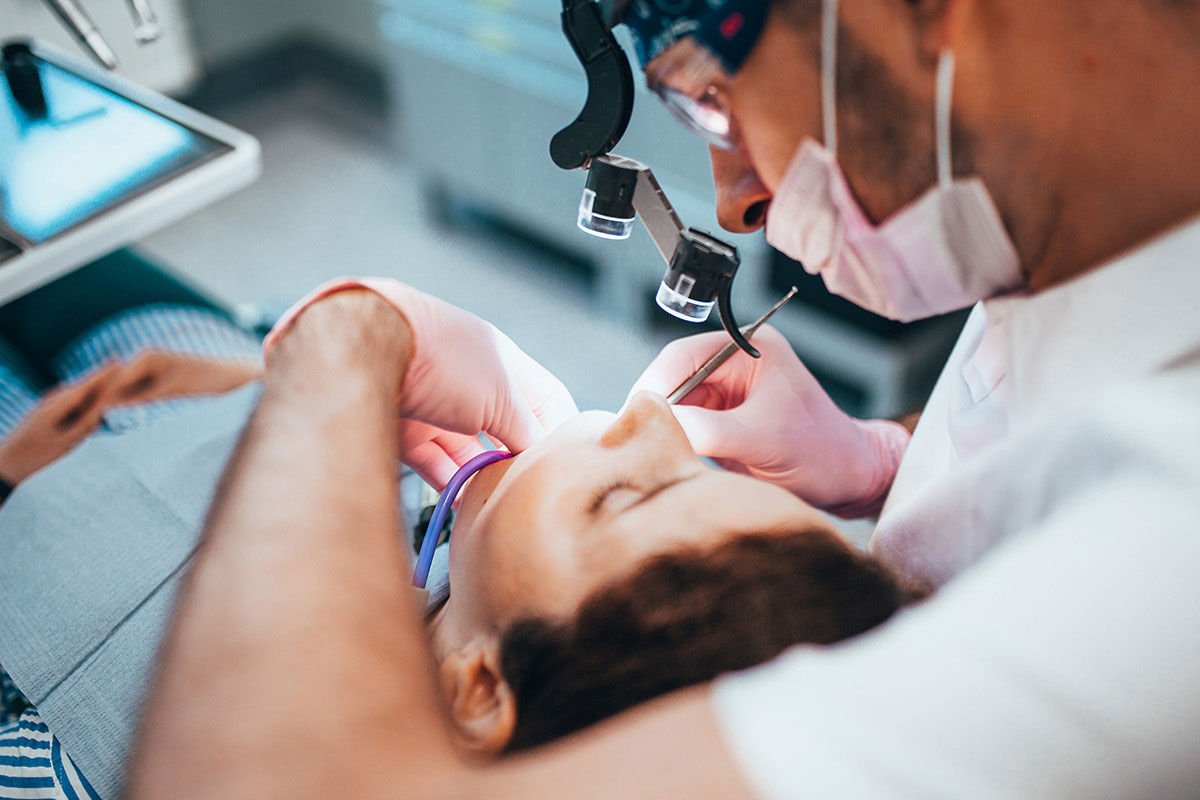
When a tooth has already received root canal treatment but inflammation remains near the tip of the root, an apicoectomy may be recommended to help preserve the natural tooth. The procedure is focused, but patients understandably want to know what the days afterward may feel like. Understanding apicoectomy recovery can help you prepare for swelling, eating, oral hygiene, activity, and follow-up. Lake Houston Endodontics provides information about endodontic surgery in Humble, TX for teeth that may need care beyond a conventional root canal.

Most apicoectomy recovery instructions focus on protecting the surgical area while the gum and underlying bone begin healing. Mild swelling, tenderness, and temporary changes around the incision may occur. However, increasing pain, significant bleeding, spreading swelling, fever, or other unusual symptoms should be reported. Knowing what to expect can make the recovery period easier to manage.

What Is an Apicoectomy?

An apicoectomy is also known as root-end surgery or root-end resection. It is an endodontic surgical procedure typically considered when inflammation or infection persists around the end of a tooth root despite previous root canal treatment.

During the procedure, the endodontist numbs the treatment area and creates a small opening in the gum near the affected root. Inflamed tissue can then be removed, along with a small portion of the root tip.

The end of the root canal may be prepared and sealed from this direction. Afterward, the gum is repositioned and closed with sutures. The objective is to treat the persistent problem while keeping the natural tooth in place.

Apicoectomy recovery begins immediately after the procedure. However, different tissues heal at different rates. The gum may improve relatively quickly, while healing within the bone continues much longer.

The First Few Hours of Apicoectomy Recovery

Local anesthetic may keep your mouth numb for several hours after treatment. During this period, avoid chewing until normal sensation returns. Otherwise, you could accidentally bite your cheek, lip, or tongue.

If sedation was used, follow all instructions regarding transportation, eating, activity, medications, and supervision. Lake Houston Endodontics also offers information about sedation dentistry for appropriate patients who need additional comfort during endodontic procedures.

A small amount of oozing can occur after surgery and should gradually decrease. Follow the instructions provided by your endodontist about gauze and pressure.

Also, avoid repeatedly pulling your lip or cheek away to inspect the surgical area. Although curiosity is understandable, stretching the tissues can place unnecessary pressure on the incision and sutures.

Swelling During Apicoectomy Recovery

Swelling may not reach its highest level immediately after the procedure. Instead, it can become more noticeable later that day or during the following day before it begins improving.

The amount of swelling varies among patients. The location of the tooth, the extent of surgery, and your individual healing response can all influence what you experience.

A cold compress placed against the outside of the face for short intervals may be recommended during the early recovery period. Follow the instructions from your surgical team regarding how and when to use it.

Mild to moderate swelling can be part of routine apicoectomy recovery. However, swelling that increases rapidly, spreads significantly, or occurs with fever or difficulty swallowing requires prompt attention.

What Should You Eat After an Apicoectomy?

Soft foods can make the first several days more comfortable. Options may include eggs, yogurt, oatmeal, pasta, soft rice, cottage cheese, mashed vegetables, and other foods that require minimal chewing.

Avoid extremely hot foods while the tissues are sensitive. In addition, stay away from hard, crunchy, sharp, or sticky items near the surgical site. Chips, popcorn, hard candy, nuts, and crusty bread can irritate the incision or become caught around sutures.

When possible, chew on the opposite side of your mouth. Meanwhile, stay hydrated and follow any specific instructions you received about drinking methods.

Tobacco use can interfere with oral healing, so follow your dental team’s recommendations regarding smoking and other tobacco products during apicoectomy recovery.

How to Manage Tenderness After Surgery

Tenderness around the incision is expected after an endodontic surgical procedure. You may notice soreness when smiling widely, touching the outside of your face, or cleaning near the area.

Use medication only according to the instructions provided by your healthcare or dental team. Medical conditions, allergies, pregnancy, and other medications can influence what pain-relief options are appropriate for you.

Apicoectomy recovery should generally move toward improvement. The area may feel more noticeable at certain times, but steadily increasing pain deserves attention.

If discomfort becomes severe, does not respond to the recommended medication plan, or returns after the area had been improving, contact the practice for guidance.

How to Keep the Surgical Area Clean

Oral hygiene remains important after surgery, but the incision needs gentle care. Continue cleaning the rest of your mouth according to your normal routine unless you receive different instructions.

Around the surgical site, follow the endodontist’s specific directions. You may be asked to modify brushing for a short period so the toothbrush does not directly disturb the sutures.

Avoid vigorous rinsing immediately after surgery unless specifically instructed otherwise. Forceful swishing and spitting can place unnecessary stress on healing tissues.

Likewise, do not use toothpicks, fingernails, or other objects to remove material near the incision. If you are uncertain about how to clean the area, contact the office for guidance.

When Can You Return to Normal Activities?

Many patients return to routine, nonstrenuous activities relatively quickly. Nevertheless, apicoectomy recovery varies depending on the procedure and individual healing response.

Plan to rest after surgery. Heavy lifting, strenuous workouts, bending, and vigorous physical activity can increase blood pressure and may contribute to throbbing or bleeding during early healing.

Your endodontist can advise when it is appropriate to resume more demanding activities. If your job involves heavy lifting, outdoor heat, or strenuous physical work, ask whether temporary modifications are recommended.

If sedation was used, follow the separate restrictions related to driving, operating equipment, making important decisions, and returning to work.

What Happens to the Sutures?

Sutures help hold the gum tissue in the appropriate position during early healing. Depending on the material used, they may dissolve naturally or need to be removed during a follow-up appointment.

The sutures can feel unusual when your tongue touches them. Nevertheless, avoid pulling or playing with them because doing so can irritate the incision.

If a suture appears loose, do not attempt to remove it yourself. Contact the dental office if you are unsure whether the area remains properly closed.

At the follow-up visit, the endodontist can examine the incision, evaluate healing, remove sutures when necessary, and answer questions about returning to normal brushing and chewing.

Benefits of a Clear Apicoectomy Recovery Plan

  • Better protection of the surgical site: Food, hygiene, and activity instructions can reduce unnecessary irritation during early healing.
  • Less uncertainty: Knowing that tenderness and swelling can occur makes normal apicoectomy recovery easier to recognize.
  • Earlier recognition of concerns: Clear warning signs help patients know when to contact the dental office.
  • More comfortable healing: Preparing soft foods, medications, and time to rest can make the first few days easier.
  • Support for natural tooth preservation: The procedure and appropriate aftercare work together toward maintaining the treated tooth.
  • Better follow-up: Scheduled examinations allow the endodontist to monitor both the gum tissue and deeper healing around the root.

How Long Does Apicoectomy Recovery Take?

There is an important difference between feeling better and complete biological healing. The gum tissue can improve relatively quickly, while the bone around the root may continue healing for several months.

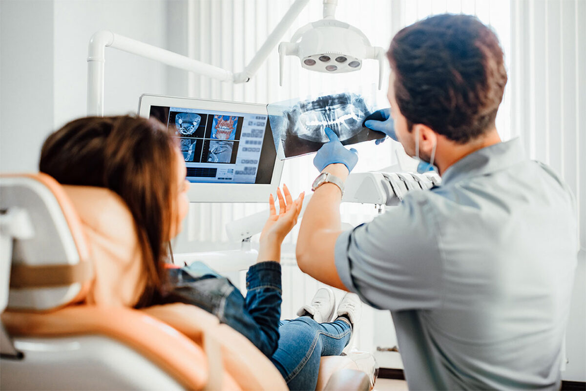
Therefore, the surgical site can look and feel comfortable before healing is complete below the surface. Follow-up dental images may be recommended so the endodontist can assess changes around the root tip.

The exact follow-up schedule depends on the tooth and the original reason for surgery. As a result, patients should attend recommended review visits even if all symptoms have disappeared.

The tooth itself may also feel slightly different during the early stage of apicoectomy recovery. For instance, biting pressure may feel tender for a period of time. This should generally improve as the supporting tissues settle.

How Apicoectomy Recovery Differs From Root Canal Recovery

A conventional root canal is performed through an opening created in the crown of the tooth. In contrast, an apicoectomy reaches the root tip through the gum and supporting tissues.

Because an apicoectomy involves an incision, gum tenderness, swelling, and sutures are more likely than they are after a routine nonsurgical root canal. Therefore, apicoectomy recovery includes soft-tissue care in addition to monitoring the tooth itself.

The procedures do share some aftercare principles. Patients should protect the treated tooth, follow medication instructions, maintain appropriate oral hygiene, and call if symptoms become significantly worse.

Some teeth may undergo endodontic retreatment before surgery is considered. In other situations, existing restorations, anatomy, or the specific location of disease can influence which approach is recommended.

When Should You Contact the Endodontist?

Routine tenderness and swelling should gradually improve. Contact the office if you experience persistent heavy bleeding, rapidly increasing swelling, fever, significant drainage, or severe pain that is not controlled by the recommended plan.

You should also call if the sutures separate, the surgical area is injured, or something about the incision concerns you. A brief evaluation may provide reassurance or identify an issue that needs attention.

Difficulty breathing or swallowing requires urgent medical evaluation. These symptoms should not be treated as routine postoperative discomfort.

Preparing for Endodontic Surgery

A little preparation can make the first day of recovery easier. Before the procedure, obtain any medications you have been instructed to use and purchase a selection of soft foods.

If the dental team recommends having a cold pack available, prepare one ahead of time. In addition, arrange transportation when sedation or the planned procedure requires it.

Provide the office with an updated medication list and mention allergies, medical conditions, pregnancy, or recent health changes. This information helps the team plan treatment and medication appropriately.

Finally, give yourself some flexibility after surgery. Avoid scheduling demanding physical activities immediately afterward. A lighter schedule can make the first stage of apicoectomy recovery more comfortable.

Frequently Asked Questions About Apicoectomy Recovery

How much swelling is normal after an apicoectomy?

Mild to moderate swelling can occur and may become more noticeable before improving. Rapidly increasing or spreading swelling should be reported promptly.

When can I eat normally?

Begin with softer foods and chew away from the surgical area. You can gradually return to your regular diet as tenderness decreases and your dental team advises.

Can I brush my teeth after apicoectomy surgery?

Yes, oral hygiene remains important. However, the surgical area may require modified brushing for a short period. Follow your specific postoperative instructions.

How long do the stitches stay in?

The timing depends on the suture material. Some sutures dissolve naturally, while others are removed during a follow-up appointment.

When should discomfort begin improving?

Postoperative tenderness should generally trend toward improvement after the early healing period. Pain that becomes increasingly severe or occurs with new swelling should be evaluated.

Give the Surgical Area Time to Recover

Apicoectomy recovery occurs gradually. The gum may look much better within a relatively short time, while bone around the root continues repairing for months. Protecting the surgical area, maintaining appropriate oral hygiene, attending follow-up appointments, and reporting unusual symptoms all support the healing process.

If you have been told you may need root-end surgery or have questions about healing after treatment, contact Lake Houston Endodontics at 19100 W Lake Houston Pkwy Ste 103 in Humble, TX. Call Us Today at (832) 777-6056 or Contact Us to schedule an appointment and learn what to expect during apicoectomy recovery.

Root Resorption After Dental Trauma: What Humble, TX Patients Should Know

A bumped, loosened, or knocked-out tooth may seem stable after the first emergency has passed, yet some dental injuries can continue changing below the surface. One concern is root resorption after dental trauma, a process in which the body begins breaking down part of a tooth’s root structure. Because the condition may be difficult to see or feel during its early stages, follow-up care after traumatic dental injuries in Humble, TX can remain important even after a tooth stops hurting.

Root resorption after dental trauma does not occur after every injury, and the term can describe several different processes. Some types begin on the outside of the root, while others begin within the tooth. The location, severity, condition of the pulp, and type of original injury all influence treatment. For patients, the key point is that an injured tooth can require monitoring long after the accident itself.

What Is Root Resorption?

Root resorption is the loss of hard tooth structure from the root. Normally, the tissues surrounding a permanent tooth help maintain the root throughout life. However, certain injuries or inflammatory processes can activate cells that begin removing dentin or cementum from the root surface.

Resorption has a normal purpose in baby teeth. As permanent teeth prepare to erupt, the roots of primary teeth gradually dissolve so those teeth can fall out. However, progressive resorption of a permanent tooth root is different and deserves evaluation.

Root resorption after dental trauma can develop when tissues that normally protect the root are damaged. Inflammation, pulp infection, and injury to the periodontal ligament can contribute to the process. Therefore, the initial appearance of the tooth does not always predict how it will heal over time.

Why Dental Trauma Can Affect the Root

A dental injury can damage much more than the visible enamel. The force of a fall, collision, sports accident, or other impact may affect the periodontal ligament, which is the thin layer of tissue between the root and surrounding bone.

The same injury may also damage the pulp, blood vessels, root surface, or developing tissues of a younger permanent tooth. In addition, the tooth can be pushed sideways, driven farther into the socket, partially displaced, or completely knocked out.

When protective root tissues are damaged, the body’s healing response can sometimes begin affecting tooth structure. If infection develops inside the pulp, inflammation can further contribute to certain forms of resorption.

The risk depends partly on the type and severity of trauma. A small enamel chip does not create the same concerns as a permanent tooth that was completely knocked out and later repositioned.

External Root Resorption After Dental Trauma

External root resorption begins on the outer surface of the root. Several patterns can occur, and they do not all progress in the same way. Some small areas may stabilize as healing occurs, while more aggressive forms can continue removing tooth structure.

Inflammatory external resorption can occur when root surface damage is combined with persistent inflammation. If the pulp becomes infected after trauma, bacteria inside the canal system can contribute to inflammation around the root.

In appropriate situations, addressing the infected pulp with root canal treatment can help remove an important source of inflammation. Nevertheless, treatment depends on the location and type of resorption rather than on one symptom alone.

Another serious pattern can involve the root becoming closely integrated with surrounding bone. Over time, normal bone remodeling can gradually replace areas of tooth structure. This process can be difficult to stop once established, which is one reason significant dental injuries deserve ongoing follow-up.

What Is Internal Root Resorption?

Internal root resorption begins from within the canal space. On dental imaging, the canal may appear enlarged or irregular in one area. In some cases, the patient has no noticeable symptoms and the finding is discovered during an examination or X-ray.

In other cases, discomfort or a visible color change can occur. Internal resorption depends on inflamed living tissue being present within part of the pulp space. Therefore, root canal treatment may be considered to remove the tissue driving the process.

If the internal defect is identified before it has significantly weakened or perforated the root, treatment may be more manageable. However, more advanced cases can be complicated, particularly if resorption has created an opening through the root wall.

Because internal and external root resorption may look similar on a traditional two-dimensional image, additional diagnostic imaging can sometimes help determine where the defect begins.

Can Root Resorption Develop Without Pain?

Yes. One of the challenges of root resorption after dental trauma is that the early stages may cause few or no symptoms. A tooth can look and feel relatively normal while changes are occurring around the root.

This is why pain alone cannot determine whether an injured tooth has healed completely. Regular follow-up after significant dental trauma allows the dental team to compare the condition of the tooth over time.

If symptoms do occur, they may include tenderness with chewing, swelling, a small pimple-like bump on the gum, drainage, tooth mobility, or discoloration. However, these signs can also appear with several other dental conditions.

Consequently, professional examination and appropriate imaging are needed to determine whether root resorption is actually present.

How an Endodontist Evaluates Root Resorption

The evaluation begins with the history of the injury. Your endodontist may ask when the accident occurred, how the tooth moved, whether it was knocked out, how it was handled, and what treatment was performed at the time.

Even an injury from many years earlier can be relevant. Therefore, mention previous falls, sports injuries, collisions, or accidents involving the tooth even if everything seemed to heal afterward.

The clinical examination may include pulp testing, gentle tapping, mobility evaluation, gum measurements, and an assessment of tooth color. Dental X-rays can show the root and surrounding bone.

When more detail is needed, three-dimensional imaging may help identify the location and extent of the resorptive area. Lake Houston Endodontics uses advanced dental technologies to assist with diagnosis and treatment planning for complex endodontic concerns.

Why Follow-Up Care Matters After Dental Trauma

Some complications do not become apparent during the initial emergency visit. The tooth may be repositioned successfully, the gum may heal, and discomfort may disappear. Nevertheless, changes inside or around the root can develop later.

Follow-up visits allow the dental team to compare current findings with earlier records. Pulp responses, root shape, bone appearance, mobility, and tooth color can all provide useful information about healing.

Root resorption after dental trauma is particularly important in this context because it may initially develop without strong symptoms. As a result, monitoring can identify a change before a patient would otherwise know that something is happening.

Patients should not skip recommended follow-up simply because the tooth feels normal. Comfort is reassuring, but examination and imaging provide information that symptoms alone cannot.

Treatment for Root Resorption After Dental Trauma

The appropriate treatment depends on whether the resorption is internal or external, how much root structure is involved, and whether pulp infection is contributing to the process.

In certain cases, monitoring may be appropriate when the change is limited and stable. In others, root canal treatment may be recommended to remove infected or inflamed pulp tissue.

If a tooth has already undergone root canal therapy and develops a new endodontic problem, endodontic retreatment may sometimes be considered. Retreatment allows the canal system to be reopened, inspected, cleaned, and resealed.

Other cases may require a surgical approach, especially when a defect is located on the root surface and cannot be adequately addressed through the inside of the tooth. Endodontic surgery can provide access to selected root problems when nonsurgical care alone is not enough.

Unfortunately, not every tooth with progressive resorption can be preserved. If too much structure has been lost or the root is no longer restorable, extraction may need to be discussed. The treatment recommendation should reflect the condition of the entire tooth and its expected long-term function.

Benefits of Early Care for Root Resorption After Dental Trauma

  • Earlier detection: Follow-up imaging may identify root changes before they create significant symptoms.
  • More potential treatment options: A limited defect may be easier to manage than advanced structural loss.
  • Better assessment of pulp health: Testing can help determine whether an injured tooth is recovering or developing pulp disease.
  • Protection for developing teeth: Children and teenagers may need specialized follow-up when permanent roots are not fully formed.
  • Clearer long-term planning: Comparing findings over time can show whether resorption is stable or progressing.
  • Support for natural tooth preservation: Timely intervention may help retain a tooth that could become more difficult to save later.

Root Resorption After a Knocked-Out Tooth

A permanent tooth that has been completely knocked out faces unique healing challenges. The periodontal ligament cells covering the root can be damaged when the tooth remains dry for too long. Aggressive handling of the root can also injure these delicate tissues.

Therefore, a knocked-out permanent tooth requires rapid professional attention. The tooth should be handled by the crown rather than the root. Appropriate storage and prompt evaluation may affect the long-term outlook.

After the tooth is repositioned and stabilized, monitoring may continue for an extended period. Root resorption after dental trauma can become apparent later, which is why repeat imaging may be recommended even when the replanted tooth initially appears stable.

Root Resorption in Children and Teenagers

Young permanent teeth require special consideration because their roots may still be developing. The treatment goals can therefore differ from those of a fully mature adult tooth.

In certain situations, preserving living pulp tissue can support continued root development. However, severe trauma can interrupt the blood supply, damage the pulp, or injure the root surface.

An endodontist considers the type of trauma, stage of root development, pulp testing, imaging, and changes over time. Therefore, repeated examinations may be especially important for younger patients.

Parents should keep recommended follow-up appointments even if the child no longer mentions the tooth. The absence of discomfort does not always provide enough information about internal healing.

What to Watch for After an Injury

After significant dental trauma, watch for increasing tenderness, swelling, drainage, a gum bump, mobility, or a change in tooth color. In addition, report any change in the bite or new discomfort when chewing.

These symptoms do not automatically mean root resorption is present. However, they are reasons to have the tooth evaluated.

Most importantly, follow the recall schedule recommended by your dental team. Root resorption after dental trauma is often understood through changes over time rather than one isolated examination.

Frequently Asked Questions About Root Resorption After Dental Trauma

Can root resorption happen without pain?

Yes. Some forms are initially painless and are discovered during follow-up imaging.

Can root resorption be stopped?

Some types can be managed when inflammation or infection is identified and treated. Other types can continue despite treatment. The outlook depends on the specific form and extent of the problem.

Does root resorption always mean the tooth must be removed?

No. Depending on the condition, monitoring, root canal treatment, retreatment, or surgery may be considered. Extraction is generally discussed when damage is too extensive for predictable restoration.

How long after an injury can root resorption appear?

The timing varies. Some changes appear relatively soon after trauma, while others may become visible months or years later.

Why might three-dimensional imaging be recommended?

Three-dimensional imaging can provide additional information about whether a defect is internal or external and how much root structure is affected.

Protect an Injured Tooth Beyond the Emergency Visit

Root resorption after dental trauma is an important reason an apparently quiet tooth may still need continued attention. The emergency visit addresses immediate problems, while follow-up care helps determine whether the pulp, root, and surrounding bone continue to heal appropriately.

If you or your child has experienced a significant dental injury, contact Lake Houston Endodontics at 19100 W Lake Houston Pkwy Ste 103 in Humble, TX. Call Us Today at (832) 777-6056 or Contact Us to schedule an evaluation for root resorption after dental trauma.

What Is a Dental Dam for Root Canal Treatment, and Why Is It Important?

Have you ever wondered why a small sheet is placed around a tooth during endodontic care? This material is called a dental dam. A dental dam for root canal treatment isolates the tooth from saliva and creates a cleaner, more controlled area for the procedure. Lake Houston Endodontics includes isolation as part of its approach to root canal treatment in Humble, TX, where careful infection control and precision are especially important when treating the inside of a tooth.

At first glance, a dental dam may seem like a minor part of the procedure. However, it performs several important jobs at once. It helps separate the opened tooth from bacteria in saliva, protects the mouth from small instruments and treatment materials, and gives the endodontist a stable working field. Therefore, understanding the purpose of a dental dam for root canal treatment can make the entire experience feel more familiar before your appointment begins.

What Is a Dental Dam?

A dental dam is a thin, flexible sheet positioned so that the tooth receiving treatment comes through a small opening. The surrounding teeth, tongue, cheeks, and much of the mouth remain behind the material. A small clamp or similar stabilizing device helps keep the dam securely positioned.

Once the dental dam is in place, the treated tooth is separated from the rest of the mouth. The endodontist can access the tooth while saliva remains outside the treatment field. This matters because saliva naturally contains bacteria, even in a healthy mouth.

During root canal treatment, the internal canal system is being cleaned and disinfected. Therefore, controlling exposure to new bacteria helps create a more predictable environment for treatment.

Why Isolation Is Important During Root Canal Treatment

A root canal procedure is different from treatment performed only on the outer surface of a tooth. During endodontic care, the tooth is opened so the endodontist can access the pulp chamber and narrow canals inside the roots.

Once those internal spaces are opened, tiny instruments and disinfecting liquids are used to remove inflamed or infected tissue and clean the canal system. Because the objective is to reduce bacteria inside the tooth, saliva control is especially valuable.

A dental dam for root canal treatment creates a physical barrier between the opened tooth and the rest of the mouth. In addition, it helps create a dry field so the endodontist can see and work more precisely.

Without effective isolation, the tongue, cheeks, moisture, and saliva would constantly move around the treatment area. Consequently, the dental dam gives the endodontist a more stable environment for delicate work inside narrow root canals.

How a Dental Dam for Root Canal Treatment Is Placed

The placement process is typically straightforward. The dental team selects the appropriate dam material and creates an opening for the tooth being treated. A small clamp may then be positioned around the tooth, and the sheet is stretched over a frame so it remains stable.

You may notice gentle pressure when the clamp is positioned. However, the setup should not cause sharp discomfort. If something pinches or feels unusual, you can let the dental team know so they can evaluate the position.

Once the dental dam for root canal treatment is secure, you can continue breathing normally through your nose. Patients sometimes worry that the sheet will interfere with breathing, but it does not cover the nose or airway.

If you have significant nasal congestion, breathing concerns, or anxiety about having material over the mouth, mention this before treatment. Communication gives the dental team an opportunity to discuss appropriate comfort options.

How the Dental Dam Helps Control Bacteria

The mouth contains many types of microorganisms. Even excellent brushing, flossing, and professional dental cleanings do not make the mouth sterile. Under normal circumstances, this is expected. During root canal treatment, however, the opened tooth needs a controlled environment.

A dental dam for root canal treatment helps prevent saliva from moving directly across the access opening. This supports the cleaning and disinfection process by limiting new contamination while the canal system is being treated.

Root canal success involves several factors, including an accurate diagnosis, cleaning, shaping, disinfection, sealing, and an appropriate permanent restoration afterward. The dental dam does not replace any of these steps. Instead, it supports them by maintaining a controlled treatment field.

How the Dental Dam Helps Protect You

Endodontic treatment involves extremely small instruments because root canals themselves can be very narrow. In addition, the procedure uses solutions intended to clean and disinfect the inside of a tooth.

A dental dam creates a physical barrier between the treatment area and the rest of your mouth. This helps reduce the chance that a small instrument or dental material could move toward the throat. It also separates the tongue and other soft tissues from the solutions used inside the tooth.

In addition, suction can be concentrated around the isolated area. Many patients find this easier because they do not have to manage as much water or movement around their tongue during treatment.

Can You Breathe Normally With a Dental Dam?

Yes. Most patients breathe normally through the nose while the dental dam is in place. The sheet isolates the tooth and mouth area, but it does not cover the nose.

Nevertheless, patients with a blocked nose, chronic breathing difficulties, a strong gag reflex, or dental anxiety should discuss those concerns before the procedure. The team can explain what to expect and determine whether adjustments are appropriate.

For patients who experience significant dental anxiety, Lake Houston Endodontics also provides information about sedation dentistry. Sedation may be an option for appropriate patients, depending on medical history and treatment needs.

It can also help to establish a simple hand signal before treatment. Raising a hand can let the dental team know that you need a brief pause. Knowing that communication remains available often makes the procedure feel less intimidating.

What Does the Dental Dam Feel Like?

The dental dam may feel unusual during the first few minutes. You will notice that the treated tooth is separated from the rest of your mouth, and the material may rest lightly around the lips.

However, many patients adjust quickly. Because the tongue is kept away from the treatment area, you do not need to concentrate on holding it still. Instead, the dental team can focus on the isolated tooth while you remain as relaxed as possible.

You may also notice pressure from the small clamp that stabilizes the dam. If that pressure becomes uncomfortable, tell the endodontic team. An adjustment may be possible without compromising isolation.

What if the Tooth Is Broken or Difficult to Isolate?

Not every tooth has the same amount of healthy structure above the gumline. Deep decay, large fillings, fractures, and previous dental procedures can make isolation more challenging.

In some situations, the endodontist may need to modify the setup or build up part of the tooth before treatment. The objective is to create a dependable seal around the working area while preserving as much healthy tooth structure as possible.

A severely damaged tooth also needs to be assessed for restorability. Root canal treatment can address disease inside the tooth, but it cannot correct every structural problem. Therefore, the remaining tooth structure and the ability to restore it are important parts of treatment planning.

Benefits of a Dental Dam for Root Canal Treatment

  • A cleaner working field: The dam helps keep saliva and oral bacteria away from the opened tooth.
  • Protection for the patient: It provides a barrier between the mouth and small endodontic instruments or treatment solutions.
  • Improved visibility: The endodontist can focus on an isolated tooth without constant movement from the tongue and cheeks.
  • Better moisture control: A dry field supports careful cleaning, inspection, and placement of treatment materials.
  • Easier fluid management: Water and cleaning solutions can be controlled within the treatment area and removed with suction.
  • A stable treatment environment: Isolation supports the detailed work required inside small root canals.

Why Isolation Is Also Important During Retreatment

A dental dam can also play an important role when a previously treated tooth requires another endodontic procedure. During endodontic retreatment, the tooth is reopened so previous filling materials can be removed and the canal system can be cleaned again.

Retreatment can involve crowns, posts, old filling materials, or difficult anatomy. As a result, maintaining a dry and stable working field becomes particularly valuable.

The dental dam separates the tooth from saliva while the endodontist carefully examines the internal anatomy. Magnification, specialized instruments, and irrigation can then be used in a controlled environment.

How the Dental Dam Fits Into the Complete Procedure

The dental dam is only one part of root canal care. First, the tooth must be properly diagnosed. The endodontist reviews your symptoms, examines the tooth, performs appropriate testing, and studies dental images.

Once treatment begins, the tooth is numbed and isolated. The canals are then located, cleaned, shaped, disinfected, dried, and filled. Afterward, an appropriate temporary restoration may be placed.

When endodontic treatment is complete, a general dentist may recommend a crown or other permanent restoration. This restoration protects the remaining tooth structure and helps maintain a dependable seal above the root canal treatment.

Each step has its own role. The dental dam for root canal treatment protects the working field during the procedure, while the permanent restoration helps protect the tooth after treatment is complete.

Frequently Asked Questions About a Dental Dam for Root Canal Treatment

Is a dental dam commonly used during root canal treatment?

Yes. Dental dam isolation is a standard part of endodontic care because it helps control saliva, protect the patient, and create a clean working environment.

Can I swallow with a dental dam in place?

Yes. Normal swallowing remains possible, and suction is used to manage fluids around the treatment area.

Does a dental dam make breathing difficult?

Most patients breathe normally through the nose. Anyone with significant nasal congestion or breathing concerns should tell the dental team before treatment.

Does dental dam placement hurt?

The dental dam itself should not be painful. Some patients notice pressure from the clamp used to stabilize it. Tell the team if the pressure becomes uncomfortable.

Why is suction alone not enough?

Saliva is continuously produced, while the tongue and cheeks move naturally around the teeth. A dental dam provides a dependable physical barrier around the tooth being treated.

A Simple Tool That Supports Precise Care

A dental dam for root canal treatment may look simple, but it supports infection control, patient protection, visibility, and moisture management throughout the procedure. By separating the tooth from the rest of the mouth, it gives the endodontist a controlled environment for precise treatment.

If you have questions about root canal treatment or what happens during an endodontic appointment, contact Lake Houston Endodontics in Humble, TX. Call Us Today at (832) 777-6056 or Contact Us to schedule an appointment and learn more about a dental dam for root canal treatment.

One Visit or Two? How Many Root Canal Appointments Will You Need?

When tooth pain is interrupting meals, sleep, or work, one of the first practical questions is often simple: how many appointments will treatment take? Many root canal appointments can be completed efficiently, but some teeth benefit from a second visit because of infection, complex anatomy, prior treatment, or the need for medication inside the canal system. Patients considering root canal treatment in Humble, TX can feel more prepared when they understand why the number of visits may differ from one tooth to another.

There is no universal rule that every root canal must be finished in one appointment or that two visits are always better. Instead, an endodontist looks at the condition of the tooth, the shape of the roots, the amount of inflammation, and what is found during treatment. Therefore, the best schedule for root canal appointments is the one that allows the tooth to be cleaned, disinfected, sealed, and protected as predictably as possible.

What Happens During Root Canal Appointments?

Root canal treatment focuses on the inside of a tooth. Beneath the enamel and dentin is a space that contains pulp tissue. When that tissue becomes severely inflamed or infected because of deep decay, a crack, trauma, or repeated dental work, the tooth may need endodontic treatment.

During root canal appointments, the tooth is numbed and isolated. Next, a small opening is created so the endodontist can reach the pulp chamber and root canals. Tiny instruments and disinfecting solutions are then used to clean and shape the internal spaces. Once the canal system is ready, it is filled and sealed with materials designed for endodontic treatment.

Although these steps may sound straightforward, teeth vary considerably. For example, a front tooth may have one relatively direct canal, while a molar may have several roots, multiple canals, curves, narrow areas, or small branches that require more time to identify and treat. In addition, a tooth with active infection may require a different approach than a tooth with inflammation that has not spread beyond the pulp.

When One Root Canal Appointment May Be Appropriate

Many teeth can be treated during a single visit when the diagnosis is clear, the canal anatomy can be managed efficiently, and the tooth can be thoroughly cleaned and sealed during the same appointment. In these situations, completing treatment in one visit can reduce the need for an additional trip and allow the tooth to move toward its permanent restoration sooner.

A one-visit approach may be considered when the endodontist can access the canals, disinfect them thoroughly, control moisture, and create a dependable seal. The decision is not simply based on how much time is available on the schedule. Instead, it depends on whether the clinical goals of treatment can be achieved during that visit.

Some patients assume that one root canal appointment means the tooth was easy to treat, while two appointments mean something went wrong. However, neither assumption is accurate. Root canal appointments are planned around the individual tooth. A complicated molar might still be completed during one appointment, while a tooth that initially appears straightforward may need another visit if unexpected anatomy is discovered.

Why Some Root Canal Appointments Are Split Into Two Visits

There are several reasons an endodontist may choose to complete treatment over two appointments. For instance, some teeth benefit from additional disinfection time. In those situations, medication may be placed inside the canals between appointments, and the tooth is protected with a temporary filling.

A second visit may also be appropriate when the tooth has unusually complex anatomy. Narrow canals, sharp curves, calcification, or difficult-to-locate spaces can require careful work under magnification. Rather than rushing to complete the procedure, the endodontist can separate treatment into stages and address each area methodically.

The condition of the tissues may matter as well. If a tooth cannot be adequately dried or there is active drainage, sealing the canal system immediately may not be the preferred approach. Therefore, root canal appointments may change based on clinical findings during the procedure.

Patient comfort is another consideration. Someone with jaw fatigue, a strong gag reflex, dental anxiety, or certain medical considerations may find a shorter appointment more manageable. Lake Houston Endodontics also offers sedation dentistry for appropriate patients who need additional support during endodontic care.

Does Two-Visit Treatment Mean the Infection Is Worse?

Not necessarily. Two root canal appointments can be recommended for several reasons, and the number of visits does not provide a simple measurement of how serious an infection is. Some teeth with significant inflammation can be treated in one visit, while other teeth with relatively few symptoms require additional time because of their anatomy.

What matters most is why the treatment plan was chosen. Your endodontist can explain whether the tooth needs additional disinfection, whether medication is being placed between visits, or whether another clinical factor is influencing the schedule.

If a temporary filling is placed between root canal appointments, protect it carefully. Avoid very hard and sticky foods on that side, continue appropriate oral hygiene, and contact the dental office if the temporary material becomes loose or comes out.

How Complex Root Anatomy Can Affect Treatment Time

Root canals are rarely identical from one person to another. Molars can contain several canals, and some canals divide, reconnect, curve, or become extremely narrow. Previous dental procedures can also change the internal anatomy. For instance, calcification can make a canal much harder to locate and enter safely.

This is one reason endodontists use specialized instruments, magnification, illumination, and advanced imaging. These technologies can help identify anatomy that may otherwise be difficult to detect. Nevertheless, precision takes time. Root canal appointments may therefore be longer or separated when a tooth requires additional attention.

In selected cases, three-dimensional imaging can help show the number, position, and shape of roots and canals before or during treatment. This information can make the procedure more targeted, particularly when traditional dental X-rays do not fully explain what is happening inside the tooth.

What About a Tooth That Already Had a Root Canal?

A tooth that received root canal treatment in the past can occasionally develop new disease months or years later. In that situation, another endodontic procedure may be considered. Patients can learn more about endodontic retreatment when a previously treated tooth becomes painful or develops new changes.

Retreatment can be more complex than an initial root canal because the endodontist may need to remove previous filling materials, work around a crown or post, and search for canals that were difficult to identify during the original procedure. Consequently, retreatment appointments may take additional time.

Even then, the number of visits varies. Some retreatment cases can be completed efficiently, while others benefit from staged care. The objective remains the same: carefully disinfect and seal the canal system while preserving the natural tooth whenever possible.

Benefits of Root Canal Appointments Planned Around Your Tooth

  • Care matched to the tooth: The treatment schedule reflects the tooth’s anatomy, infection level, and individual needs rather than following a rigid formula.
  • Thorough disinfection: Additional time can be used when medication or further cleaning is appropriate before permanent sealing.
  • Greater patient comfort: Longer procedures can sometimes be divided when shorter visits are easier to manage.
  • Careful management of complex anatomy: Narrow, curved, calcified, or hidden canals can receive the attention they require.
  • Clear treatment progression: Patients can understand when the endodontic procedure is complete and when restorative care should follow.
  • Support for natural tooth preservation: Each appointment focuses on creating favorable conditions for long-term function.

What to Expect Between Root Canal Appointments

If your treatment is divided into two visits, the tooth is usually closed with a temporary filling. It may feel mildly tender afterward, particularly if it was painful before treatment. This pressure sensitivity often comes from the tissues around the root.

Follow the instructions provided by the practice. Take medications only as directed and avoid putting unnecessary chewing pressure on the temporary restoration. Meanwhile, brushing remains important, although you may need to be gentle if nearby gum tissue is sensitive.

Most importantly, do not skip the second appointment because the tooth suddenly feels better. Relief after an initial visit does not mean the root canal system is permanently sealed. Treatment should be completed as recommended to reduce the risk of new contamination.

What Happens After Root Canal Treatment Is Complete?

Once the root canal is finished, the tooth may still need protection above the gumline. Depending on its location and how much natural structure remains, your general dentist may recommend a crown or another permanent restoration.

A temporary filling is not designed to function indefinitely. Therefore, arrange the permanent restoration within the timeframe recommended by your dental team. A durable restoration helps protect the remaining tooth structure and maintains a strong seal over the endodontically treated tooth.

Afterward, continue routine dental care. Daily brushing and flossing, professional cleanings, examinations, and appropriate dental imaging can help protect the treated tooth and surrounding teeth.

Frequently Asked Questions About Root Canal Appointments

Can a root canal really be completed in one visit?

Yes. Many root canals can be completed during one appointment when the tooth can be thoroughly cleaned, disinfected, dried, and sealed during that visit.

Why would medication be placed inside the tooth between visits?

Medication may be used when additional disinfection is helpful before the canals are permanently filled. The tooth is usually protected by a temporary restoration between appointments.

How long does a root canal appointment take?

Appointment length varies depending on tooth type, anatomy, infection, and complexity. A molar with several canals can require more time than a front tooth.

Is one visit better than two visits?

Neither approach is automatically better for every tooth. The appropriate schedule depends on the clinical situation and whether treatment can be completed thoroughly and predictably.

Can I go back to work after my appointment?

Many patients return to routine activities after treatment. However, patients who receive sedation should follow the transportation and activity instructions provided by the office.

Know What Your Treatment Plan Involves

The number of root canal appointments you need is determined by what gives the tooth the best opportunity to be cleaned, sealed, restored, and preserved. One appointment may be appropriate for one tooth, while a carefully planned second visit may be better for another.

If you have tooth pain or have been told you may need endodontic care, contact Lake Houston Endodontics in Humble, TX. Call Us Today at (832) 777-6056 or Contact Us to schedule an appointment and learn what your root canal appointments may involve.

Why You Need a Crown After Root Canal Treatment: Protecting Your Tooth for the Long Term

A root canal can relieve deep tooth pain and remove infection from inside a tooth, but the work is not always finished when the endodontic appointment ends. Many back teeth need a crown after root canal treatment to protect the remaining structure and restore dependable chewing. Lake Houston Endodontics explains the internal treatment process on its root canal treatment page, including the importance of returning to a general dentist for the permanent restoration.

The root canal and the crown perform different jobs. Root canal treatment cleans and seals the internal canal system. The crown covers and reinforces the part of the tooth above the gum. Together, these treatments can help a natural tooth remain comfortable and functional. Delaying a crown after root canal treatment may leave the tooth vulnerable to fracture, leakage, and new contamination.

What Changes After a Root Canal?

Before treatment, a tooth may already be weakened by deep decay, a large filling, a crack, or repeated dental procedures. To reach the infected or inflamed pulp, the endodontist also creates a controlled opening through the chewing surface or the back of the tooth. Although the opening is kept as conservative as possible, the tooth may have less remaining structure than a healthy, untouched tooth.

A root canal does not make a tooth hollow, and the tooth does not become useless. However, the combination of existing damage and lost structure can increase the risk of fracture. Back teeth are exposed to strong chewing forces, so they often need greater coverage than a small filling can provide.

A crown after root canal treatment wraps around the visible portion of the tooth and helps distribute pressure. The exact restoration depends on the tooth, location, bite, remaining structure, and treatment plan from the general dentist.

Why a Temporary Filling Is Not the Permanent Answer

At the end of root canal treatment, a temporary filling may be placed in the access opening. This material seals the tooth between appointments and allows the endodontist to confirm that symptoms are improving. However, a temporary filling is designed for short-term protection.

Temporary materials can wear, crack, or loosen. If the seal breaks, saliva and bacteria may reach the internal space. In addition, the untreated outer tooth remains exposed to chewing forces. Therefore, a patient should not assume that the temporary filling provides the same protection as a crown after root canal treatment.

Follow the endodontist’s instructions about when to contact the general dentist. In many cases, the restorative appointment should be arranged promptly. Call sooner if the temporary filling comes out, the tooth feels loose, or a section breaks.

Which Teeth Commonly Need Crowns?

Molars and premolars commonly need crowns because they perform most of the heavy chewing. Their broad surfaces and multiple cusps experience forces from different directions. A large filling or weakened cusp can flex under pressure and eventually fracture.

Front teeth may not always require a full crown after root canal treatment. If a front tooth has minimal structural damage and a small access opening, a bonded filling may sometimes be appropriate. However, a front tooth with a large restoration, fracture, discoloration, or extensive loss of structure may still need a crown or another protective restoration.

There is no universal rule that fits every tooth. The general dentist evaluates the amount of healthy structure, appearance, bite, and long-term function. The endodontist can also share information about cracks, internal anatomy, and the treatment performed.

How a Crown Helps Prevent Fracture

Natural tooth enamel is strong, but a tooth with a large filling can behave like a shell with thinner walls. When you bite, separate cusps may flex slightly. Repeated movement can create or extend cracks. Eventually, a cusp may break, or a fracture may travel below the gum.

A crown after root canal treatment holds the remaining tooth together and spreads chewing forces over a broader area. It cannot make the tooth indestructible, and it cannot stop every crack from progressing. Still, it offers much stronger coverage than a temporary filling alone.

If a tooth already has signs of a crack, timing becomes especially important. Lake Houston Endodontics evaluates cracked teeth in Humble, TX and can explain whether the pulp needs treatment. After endodontic care, the general dentist restores the tooth according to the crack pattern and remaining structure.

How a Crown Protects the Root Canal Seal

Successful endodontic treatment depends on cleaning and sealing the canal system. However, the top of the tooth also needs a dependable seal. If a restoration leaks, bacteria from the mouth may move toward the treated canals.

A well-designed crown or other permanent restoration helps close the tooth above the gum. The dentist also rebuilds missing structure under the crown with a core material. In selected cases, a post may be placed inside part of the root to help retain the core, but a post does not strengthen the root itself. It is used only when there is not enough coronal structure to hold the restoration.

Good fit, appropriate bite adjustment, and daily hygiene all influence the long-term seal. A crown after root canal treatment still needs brushing, flossing, and regular dental examinations.

What Can Happen if the Crown Is Delayed?

Some patients feel much better after the root canal and postpone the restorative visit because the pain is gone. Unfortunately, comfort does not mean the tooth is fully protected. A temporary filling can remain in place longer than intended, and the surrounding tooth can fracture without much warning.

A small chip may be repairable. However, a deep fracture can extend below the gum or into the root, making the tooth difficult or impossible to restore. In addition, leakage can allow new contamination and may lead to the need for endodontic retreatment.

Do not chew ice, hard candy, popcorn kernels, or other very firm foods on the treated tooth while waiting for the crown. Use the opposite side when possible, and arrange the permanent restoration within the timeframe recommended by the dental team.

Benefits of a Crown After Root Canal Treatment

  • Greater fracture resistance: A crown helps hold weakened cusps together and distribute chewing pressure.
  • Protection of the internal seal: The permanent restoration reduces the chance of saliva and bacteria entering the treated tooth.
  • Restored chewing function: A properly shaped crown helps the tooth contact neighboring and opposing teeth appropriately.
  • Improved appearance: Tooth-colored materials can restore the shape and color of a damaged tooth.
  • Support for long-term tooth preservation: A crown after root canal treatment helps protect the investment made in saving the natural tooth.
  • Replacement of lost structure: The restoration rebuilds areas weakened by decay, old fillings, fractures, and the access opening.

What to Expect During Crown Treatment

The general dentist first evaluates the tooth and removes temporary material as needed. A core buildup may be placed to replace missing structure and create a stable foundation. The tooth is then shaped so the crown can fit over it without feeling bulky.

An impression or digital scan records the prepared tooth and surrounding bite. Depending on the office and material, the crown may be made during the visit or fabricated by a dental laboratory. A temporary crown may protect the tooth while the final restoration is being completed.

At the delivery appointment, the dentist checks fit, contact with neighboring teeth, appearance, and bite. Small adjustments may be made before the crown is bonded or cemented. Tell the dentist if the tooth feels high when you close because an uneven bite can create pressure tenderness.

Crown Materials and Treatment Choices

Crowns can be made from several materials, including ceramics, zirconia, porcelain fused to metal, and metal alloys. Each option has different strengths, appearance, thickness requirements, and wear characteristics. The best material depends on the tooth’s location, the patient’s bite, cosmetic goals, and the dentist’s recommendation.

A strong ceramic may be selected for a back tooth that handles heavy force. A highly aesthetic material may be considered for a visible front tooth. Patients who grind or clench may need special planning and a protective nightguard after treatment.

The endodontist usually does not fabricate the final crown. Instead, the endodontist treats the root canal system and communicates relevant findings to the restorative dentist. This teamwork helps connect internal health with external protection.

How to Care for the Crown and Treated Tooth

Brush the crown twice daily with fluoride toothpaste and clean between the teeth with floss or another recommended tool. The crown itself cannot develop decay, but the natural tooth at its margin can. Keeping the gumline clean helps protect the seal and surrounding tissues.

Avoid using teeth as tools to open packages or hold objects. Also, do not chew ice or very hard items. These habits can damage crowns and natural teeth. If you grind at night, ask the general dentist whether a nightguard would help reduce stress.

Attend regular checkups so the dentist can examine the crown margin, bite, gum health, and root area. Contact the office between visits if the crown feels loose, food repeatedly catches around it, or the tooth develops new pain or swelling.

When New Symptoms Need an Endodontic Evaluation

A root canal treated tooth does not contain the same responsive pulp tissue, but the ligament and bone around the root can still feel pressure and inflammation. New tenderness may come from an uneven bite, a crack, gum disease, leakage, or renewed infection.

If symptoms appear after the crown is placed, start by calling the dental office. An examination can determine whether the issue is restorative or endodontic. Persistent swelling, a gum bump, drainage, or a change near the root may lead to a referral for specialty evaluation.

Do not assume that a crown after root canal treatment guarantees the tooth can never develop another problem. It offers important protection, but long-term success also depends on the original diagnosis, quality of treatment, structural condition, hygiene, and ongoing dental care.

Frequently Asked Questions About a Crown After Root Canal Treatment

Does every root canal treated tooth need a crown?

No. Many back teeth do, while some front teeth may be restored with a bonded filling. The decision depends on the location, remaining structure, bite, and existing damage.

How soon should I get the crown?

Follow the timeframe given by the endodontist and general dentist. The permanent restoration is usually arranged promptly because a temporary filling is not designed for long-term protection.

Can I chew normally before the crown is placed?

Use caution. Avoid hard, sticky, and crunchy foods on the treated tooth, especially while a temporary filling is present. Chew on the opposite side when possible.

Why do I need a crown if the nerve is gone?

The crown is not placed to treat the nerve. It protects the remaining tooth structure, restores chewing shape, and helps seal the tooth above the root canal filling.

Can a crowned root canal tooth still crack?

Yes. A crown reduces risk but cannot prevent every fracture. Existing cracks, heavy grinding, limited tooth structure, and biting hard objects can affect the outlook.

Give the Treated Tooth Lasting Protection

A crown after root canal treatment is often the part of care that allows a comfortable tooth to return to reliable daily function. The root canal addresses infection inside the tooth, while the permanent restoration helps prevent fracture and leakage from above. Completing both parts on schedule gives the tooth a stronger foundation for the years ahead.

For root canal care and guidance about protecting a treated tooth, contact Lake Houston Endodontics in Humble, TX. Call Us Today at (832) 777-6056 or contact the practice online to schedule an appointment.

Calcified Root Canals in Humble, TX: Why Canals Narrow and How Endodontists Treat Them

Have you been told that a root canal is difficult to find, unusually narrow, or partly blocked? The inside of a tooth can gradually form additional hard tissue, making the canal space smaller and harder to see. These calcified root canals often require advanced imaging, magnification, and specialized instruments. Patients can learn more about root canal treatment in Humble, TX when complex anatomy makes specialty care especially valuable.

A calcified canal does not automatically mean that a tooth is infected, and it does not always require treatment. However, when the tooth develops pain, swelling, or disease around the root, the narrowed pathway can make treatment more challenging. An endodontist evaluates whether treatment is needed and how calcified root canals can be approached while preserving as much tooth structure as possible.

What Does Root Canal Calcification Mean?

Inside every tooth is a pulp chamber and one or more root canals. These spaces contain soft tissue during the life of the tooth. Over time, the tooth may deposit additional dentin along the walls. As that hard tissue builds, the visible space becomes narrower.

This process is often called calcification, canal obliteration, or pulp canal narrowing. The canal is not usually filled with a loose stone or removable plug. Instead, the walls have gradually thickened inward. Therefore, locating calcified root canals requires careful navigation through natural tooth structure.

A canal may be partially narrowed or appear almost completely closed on an X-ray. Even when the space is difficult to see, a tiny pathway may remain. The challenge is finding that path without removing more tooth structure than necessary.

Why Do Calcified Root Canals Develop?

Calcification can occur as part of normal aging. Teeth continue to form dentin over time, so the pulp chamber of an older tooth may look smaller than it did years earlier. Repeated irritation can also accelerate the process.

A history of dental trauma is another common factor. A tooth that was hit, pushed, or loosened may respond by producing hard tissue inside. The crown can become more yellow as the internal space narrows. This may happen even when the original injury occurred many years ago.

Large fillings, crowns, deep decay, or repeated procedures may also stimulate the tooth to form protective dentin. In addition, some teeth naturally have very narrow canals. The endodontist considers the patient’s age, history, restorations, symptoms, and imaging before deciding why the canal appears calcified.

Do Calcified Root Canals Cause Symptoms?

Many calcified root canals cause no symptoms. A dentist may notice the change on a routine X-ray, or a tooth may appear slightly more yellow than neighboring teeth. If the pulp remains healthy and there are no signs of disease, treatment may not be necessary.

Problems arise when the remaining pulp becomes inflamed or infected. The tooth may become sensitive to biting, ache without a trigger, develop swelling, or show a dark area near the root on an image. In some cases, the tooth no longer responds normally to cold testing because the thick dentin insulates the remaining tissue.

Diagnosis can be more complicated because reduced temperature response does not always mean the pulp is nonvital. Calcified root canals may simply make the stimulus harder to feel. The endodontist combines several tests and compares the tooth with nearby teeth before recommending treatment.

How Calcified Root Canals Are Diagnosed

The examination begins with the history of the tooth. Previous trauma, a color change, a large restoration, or an old injury may explain why the canal narrowed. The endodontist also asks about biting pain, swelling, temperature sensitivity, and how long the symptoms have been present.

Digital X-rays show the size and general direction of the pulp chamber and roots. If the canal is not visible on a two-dimensional image, advanced dental technology may provide more detail. A focused CBCT scan can sometimes show the remaining pathway and its relationship to the outer root surface.

Pulp testing, tapping, palpation, gum measurements, and bite testing may also be used. The purpose is to answer two separate questions: Does the tooth need treatment, and if it does, how can the canal be reached safely?

Why Specialty Training Matters

Calcified root canals can be difficult because the normal landmarks inside the tooth may be less obvious. Searching too aggressively can remove unnecessary dentin, weaken the tooth, or create a perforation. A perforation is an unintended opening from the root canal space to the outside of the tooth.

Endodontists focus on diagnosing and treating problems inside teeth. They use detailed knowledge of tooth anatomy, high magnification, illumination, and small instruments to approach narrow canals. This specialty training is particularly useful when a general dentist identifies a canal that may be difficult to locate.

Referral does not mean the tooth is hopeless. Instead, it often means the tooth deserves a more specialized approach. The endodontist will still discuss limitations, alternatives, and the expected outlook before treatment begins.

How Dental Microscopes Help

A dental operating microscope enlarges the treatment area and adds bright, focused light. Calcified root canals may be represented by subtle color changes, tiny grooves, or small differences in the floor of the pulp chamber. These details can be difficult to recognize without magnification.

The microscope also helps the endodontist work conservatively. Rather than opening a wide area and hoping to find the canal, the clinician can follow small anatomical clues. This supports preservation of the surrounding dentin, which is important for the strength of the tooth.

Magnification does not make every calcified canal easy, but it improves visibility and control. It is often combined with ultrasonic instruments and three-dimensional imaging.

Ultrasonic Instruments and Guided Access

Ultrasonic tips vibrate at a high frequency and can remove small amounts of hard tissue with precision. During treatment of calcified root canals, they may be used to uncover an opening or refine access while the area is viewed under a microscope.

Very small hand instruments can then explore the pathway. The endodontist uses a gentle, controlled technique because narrow canals can curve or change direction. Lubricating materials and electronic measurement tools may support navigation.

In highly complex cases, information from a CBCT scan can be used to plan a guided access path. Not every calcified tooth is suitable for the same technique. The best approach depends on the location of the narrowing, the amount of remaining tooth structure, and the experience of the treating clinician.

Benefits of Treating Calcified Root Canals

  • Removal of infected tissue: When disease is present, treatment cleans the remaining canal space and addresses the source inside the tooth.
  • Preservation of the natural tooth: Successful treatment of calcified root canals may avoid extraction and replacement.
  • Relief from pain or pressure: Addressing inflammation can improve chewing comfort and reduce spontaneous aching.
  • Use of advanced visualization: Microscopes and focused imaging help the endodontist locate narrow anatomy more precisely.
  • Conservative access: Specialized techniques aim to preserve as much healthy dentin as possible.
  • Better treatment planning: A detailed evaluation clarifies whether nonsurgical care, surgery, monitoring, or another option is appropriate.

What Happens if the Canal Cannot Be Reached?

Despite careful treatment, some calcified root canals cannot be negotiated safely from the top of the tooth. Continuing to search may create more risk than benefit. When this happens, the endodontist stops and reassesses rather than removing excessive tooth structure.

Depending on the diagnosis, endodontic surgery may provide access to the root tip from outside the tooth. An apicoectomy can remove inflamed tissue near the end of the root and seal the canal from that direction. Surgery is not the automatic answer, but it may be considered when nonsurgical access is limited.

In other cases, monitoring may be appropriate if there are no symptoms or signs of active disease. Extraction may be discussed when the tooth is severely cracked, cannot be restored, or has an unfavorable long-term outlook. The recommendation should reflect the entire tooth, not only the fact that the canal is calcified.

Recovery After Treatment

A treated tooth may feel tender for several days, especially if it was sore before the appointment. Pressure sensitivity usually comes from the ligament around the root rather than the removed pulp tissue. Follow the medication and care instructions provided by the office.

Avoid chewing hard foods on the tooth while a temporary filling is present. Calcified root canals often occur in teeth with a history of trauma, large restorations, or other structural changes, so protecting the tooth is important. Return to the general dentist for the recommended crown or permanent restoration.

Call the office if pain becomes severe, swelling increases, the bite feels high, or the temporary filling comes out. Steady improvement is expected, while worsening symptoms deserve evaluation.

Can Calcification Be Prevented?

Normal age-related dentin formation cannot be prevented. Likewise, a tooth may calcify as part of its healing response after an injury. However, reducing avoidable trauma and treating decay early can help protect the pulp from repeated irritation.

Wear a mouthguard for contact sports and avoid chewing ice, hard candy, or other objects that can damage teeth. If a tooth is injured, seek an examination even when it does not hurt. Early records create a baseline that can be compared with future images.

Maintain regular dental visits so changes in tooth color, canal size, or root health can be monitored. A calcified tooth may remain stable for years, but follow-up makes it easier to identify a problem if one develops.

Frequently Asked Questions About Calcified Root Canals

Does a calcified canal always need a root canal?

No. A tooth may have a narrowed canal and remain healthy. Treatment is recommended when symptoms, testing, or imaging show that the pulp or tissues around the root require care.

Can a calcified tooth still hurt?

Yes. Remaining pulp tissue can become inflamed or infected, and the tissues around the root can become tender. The tooth may hurt with biting or develop swelling even when the canal is difficult to see.

Why is the tooth more yellow?

Additional dentin inside the tooth can make the crown look darker or more yellow. A color change should be evaluated, especially when there is a history of trauma.

Are calcified root canals harder to treat?

They can be more challenging because the pathway is narrow and landmarks may be less visible. Magnification, illumination, ultrasonics, and three-dimensional imaging can support treatment.

What if the canal cannot be found?

The endodontist may discuss monitoring, surgery, or another treatment based on symptoms, restorability, and the location of disease. Continuing to remove tooth structure without a safe path may not be advisable.

Protect a Tooth With Complex Anatomy

Calcified root canals require patience, detailed diagnosis, and a conservative plan. The important question is not simply whether the canal looks narrow. Instead, the endodontist determines whether the tooth is diseased, whether the pathway can be reached safely, and which option offers the most predictable way to preserve function.

If you have been referred for a difficult or calcified canal, contact Lake Houston Endodontics in Humble, TX. Call Us Today at (832) 777-6056 or contact the practice online to schedule an evaluation for calcified root canals.

CBCT Imaging for Root Canal Treatment: How 3D Scans Reveal Hidden Problems

A standard dental X-ray can show a great deal, but some tooth problems hide behind overlapping roots, dense bone, or complex internal anatomy. In selected cases, CBCT imaging for root canal treatment gives an endodontist a three-dimensional view that can make diagnosis and planning more precise. Lake Houston Endodontics uses advanced dental technologies in Humble, TX to investigate difficult symptoms and plan specialized care.

A CBCT scan is not needed for every toothache or every root canal. Instead, it is used when the additional information may change the diagnosis, reveal anatomy that cannot be seen clearly on two-dimensional images, or help the endodontist choose between treatment options. Understanding what the scan does can help patients feel more confident when CBCT imaging for root canal treatment is recommended.

What Is a Dental CBCT Scan?

CBCT stands for cone beam computed tomography. The scanner moves around the patient’s head and captures many small images from different angles. Computer software then combines those images into a three-dimensional view of the teeth, roots, jawbone, and nearby structures.

The resulting data can be viewed in thin slices from several directions. An endodontist may rotate the image, enlarge a particular root, or examine the area from the side, front, and top. This flexibility is valuable because root canal systems are three-dimensional, even though traditional dental X-rays flatten the area into a two-dimensional picture.

CBCT imaging for root canal treatment is focused on diagnosis and planning. It does not replace a clinical examination, pulp testing, bite testing, or standard digital X-rays. Instead, it adds another layer of information when the basic findings leave an important question unanswered.

How CBCT Differs From a Standard Dental X-Ray

A standard dental X-ray is quick, useful, and appropriate for many endodontic evaluations. It can show decay, large restorations, bone changes, previous root canal fillings, and the general shape of roots. However, structures can overlap. A root positioned behind another root may be difficult to separate, and a small area of bone loss may not be visible from the angle used.

CBCT imaging for root canal treatment reduces the problem of overlap by displaying the area in three dimensions. For example, the endodontist may be able to see whether a dark area is associated with one root or another. The scan may also show that a root curves in a direction that was hidden on the standard image.

Still, more imaging is not always better. A standard X-ray may already provide everything needed for a straightforward case. The endodontist chooses CBCT when the expected diagnostic value justifies the scan.

Finding Extra or Unusual Root Canals

Teeth do not all follow a simple blueprint. A molar may have an extra canal, a canal may divide and reconnect, or roots may curve sharply. These variations can be difficult to identify on a flat image. If infected tissue remains in an untreated canal, symptoms or disease may continue after treatment.

CBCT imaging for root canal treatment can help map unusual anatomy before the procedure begins. This gives the endodontist a clearer idea of where to look and how the canals may travel through the root. Magnification and illumination are then used during treatment to locate and clean the actual openings.

The scan does not perform the treatment, and it cannot guarantee that every microscopic branch will be visible. However, a three-dimensional roadmap can make a complex case more understandable and support a more efficient plan.

Evaluating a Tooth That Still Hurts After a Root Canal

A previously treated tooth may become painful months or years later. Causes can include a missed canal, leakage from a restoration, new decay, a fracture, or persistent inflammation around a root. Because root filling materials and crowns can make standard images harder to interpret, additional views may be useful.

CBCT imaging for root canal treatment can show the location and size of bone changes around individual roots. It may also reveal untreated anatomy or a problem that affects only one side of a tooth. These details help the endodontist determine whether endodontic retreatment, surgery, monitoring, or another option deserves consideration.

Not every source of pain appears on a scan. Bite problems, muscle pain, early cracks, and some inflammatory conditions may require clinical testing and careful history. Therefore, imaging is interpreted as part of the whole examination rather than treated as the only answer.

Planning Endodontic Surgery

When inflammation remains near a root tip after nonsurgical treatment, an apicoectomy or another form of endodontic surgery may be considered. Before surgery, the endodontist needs to understand the root’s position, nearby bone, and relationship to surrounding anatomy.

CBCT imaging for root canal treatment and surgery can help identify which root is involved and where the problem is located. It may also show the thickness of bone over the root or the proximity of nearby structures. This information supports a more targeted surgical approach.

Patients can learn more about endodontic surgery in Humble, TX when nonsurgical treatment alone cannot address the problem. The recommendation depends on the tooth, restorability, and expected long-term outlook.

Can CBCT Find a Cracked Tooth?

Cracks are among the most challenging dental problems to diagnose. A large fracture or the bone pattern associated with a root fracture may be visible on CBCT. However, very fine cracks can still be too small to appear clearly. Restorations and root filling materials may also create visual artifacts that make interpretation harder.

For this reason, CBCT imaging for root canal treatment is only one part of a cracked tooth evaluation. Bite testing, magnification, transillumination, gum measurements, and symptom patterns may provide important clues. Pain when releasing a bite, isolated deep gum probing, or a recurring gum bump can influence the diagnosis even if the crack itself is not visible.

A scan can still be valuable because it may rule in or rule out other causes. For example, it may show a missed canal or localized bone loss that changes the treatment discussion.

Benefits of CBCT Imaging for Root Canal Treatment

  • Three-dimensional detail: The scan allows roots and surrounding bone to be viewed from multiple directions.
  • Improved anatomy mapping: CBCT imaging for root canal treatment may reveal extra canals, unusual curves, or root variations.
  • More focused retreatment planning: The endodontist can identify which root or region may be associated with persistent disease.
  • Better surgical preparation: A scan can clarify the location of a root tip and nearby structures before endodontic surgery.
  • Support for difficult diagnosis: CBCT may provide useful information when symptoms continue but standard images are inconclusive.
  • Clearer patient communication: Three-dimensional views can help patients understand why a treatment option is being recommended.

What the Scan Experience Is Like

CBCT imaging is generally quick. You may stand or sit while the machine is positioned around your head. The team asks you to remain still, and the scanner rotates around the area. Nothing is placed deep inside the mouth, although positioning supports may be used to help you stay steady.

The scan itself does not hurt. After the image is captured, the endodontist reviews the data on a computer. In many cases, the most relevant views can be shown during the consultation so you can see the roots and surrounding bone.

Metal jewelry, removable appliances, or other objects near the head may interfere with the image. Follow the team’s instructions about removing earrings, glasses, or removable dental items before the scan.

How Imaging Is Selected Thoughtfully

CBCT imaging for root canal treatment is ordered when the expected benefit is meaningful. The field of view can often be limited to the region being evaluated, rather than scanning a larger area than necessary. The dental team also considers whether previous imaging is available and whether a standard X-ray can answer the question.

Patients should tell the office if they are pregnant or may be pregnant before imaging. They should also mention recent scans so the endodontist can determine whether those records may be useful. If another dental office has already taken a CBCT scan, ask whether the file can be transferred for review.

It is reasonable to ask why the scan is recommended. A clear answer might include locating an extra canal, evaluating a root fracture, measuring a bone lesion, or planning surgery. The reason should be connected to a specific diagnostic or treatment decision.

How CBCT Works With Microscopes and Digital X-Rays

Endodontic technology is most effective when tools complement one another. Digital X-rays provide efficient two-dimensional views. CBCT offers three-dimensional context. Meanwhile, a dental microscope gives high magnification and illumination inside the tooth during treatment.

For example, CBCT imaging for root canal treatment may suggest that a narrow extra canal is present. The microscope then helps the endodontist search for the canal opening. Ultrasonic instruments may be used to remove small amounts of calcified material while preserving surrounding tooth structure.

No technology replaces training and judgment. The endodontist must decide which finding matters, whether it matches the symptoms, and whether treatment can improve the tooth’s outlook.

Frequently Asked Questions About CBCT Imaging for Root Canal Treatment

Does every root canal need a CBCT scan?

No. Many root canals can be diagnosed and treated with an examination and standard digital X-rays. CBCT is used when three-dimensional information may improve diagnosis or planning.

Can a CBCT scan show infection?

It can show changes in the bone that may be associated with inflammation or infection. The endodontist combines those findings with symptoms, testing, and the condition of the tooth.

Will CBCT always show a crack?

No. Some cracks are too fine to be seen directly, and metal or filling materials can affect the image. Clinical tests remain important in a cracked tooth evaluation.

Is the scan uncomfortable?

The scan is generally quick and painless. The main requirement is remaining still while the machine rotates around the head.

Can an old CBCT scan be used?

Possibly. The value depends on when it was taken, the area included, image quality, and whether the tooth has changed since then. The endodontist can review the file and decide whether new imaging is needed.

See the Full Picture Before Treatment

CBCT imaging for root canal treatment can reveal details that are hidden by overlapping anatomy on a standard X-ray. When used selectively, it helps an endodontist investigate complex pain, plan retreatment, evaluate surgery, and explain findings more clearly. The scan is most useful when it answers a specific question and is interpreted alongside a complete clinical examination.

For advanced endodontic diagnosis in Humble, TX, contact Lake Houston Endodontics. Call Us Today at (832) 777-6056 or contact the practice online to schedule an appointment and learn whether CBCT imaging for root canal treatment may be appropriate for your tooth.

Root Canal Treatment During Pregnancy in Humble, TX: Safety, Timing, and Comfort

A sudden toothache can be stressful at any time, but it may feel especially complicated when you are expecting a baby. Many patients wonder whether they should delay care, avoid dental X-rays, or try to manage the pain until after delivery. In reality, untreated infection can create its own concerns, so a timely evaluation is important. Lake Houston Endodontics provides root canal treatment in Humble, TX for patients who need focused care to relieve pain and preserve a natural tooth.

Root canal treatment during pregnancy is planned with your comfort, stage of pregnancy, medical history, and dental condition in mind. Your endodontist can also coordinate with your obstetric provider when additional guidance is helpful. The goal is not to rush into treatment or postpone necessary care without a reason. Instead, the goal is to make a thoughtful decision based on the health of the tooth and the needs of the patient.

Why Tooth Pain Should Be Evaluated During Pregnancy

Pregnancy brings many physical changes, and dental symptoms can sometimes be easy to dismiss as one more temporary discomfort. However, a tooth that throbs, keeps you awake, reacts strongly to heat, or causes swelling may have inflammation or infection inside it. These problems usually do not resolve simply because a patient avoids chewing on the area.

When the pulp inside a tooth becomes severely inflamed or infected, the source is enclosed within the hard structure of the tooth. Therefore, brushing more often or taking medication may reduce symptoms for a short time, but those steps do not remove damaged tissue from the root canal system. An examination can determine whether the tooth needs monitoring, restorative care, or root canal treatment during pregnancy.

Prompt evaluation is also useful because dental pain can affect eating, sleeping, hydration, and daily comfort. In addition, swelling can worsen quickly. Contact a dental professional promptly if you notice facial swelling, drainage, fever, a gum bump, a bad taste, or difficulty swallowing. Severe or spreading symptoms need urgent attention.

Is Root Canal Treatment During Pregnancy Possible?

Yes, root canal treatment during pregnancy can be performed when it is needed. Dental care is individualized, so the timing and details depend on the patient’s health, the condition of the tooth, and the urgency of the symptoms. A painful or infected tooth may require care during any trimester, especially when delaying treatment could allow the problem to become more difficult to manage.

For nonurgent procedures, many patients find the middle portion of pregnancy more comfortable because early nausea may have improved and lying back may still be manageable. However, a dental emergency does not always follow an ideal calendar. If a tooth is causing severe pain or infection, the dental team can discuss how to provide appropriate care rather than asking the patient to endure symptoms for months.

Before the appointment, share your due date, pregnancy history, allergies, current medications, and any recommendations from your obstetric provider. Also mention complications such as high blood pressure, bleeding concerns, or a pregnancy that requires special monitoring. Complete information helps the endodontist plan root canal treatment during pregnancy more carefully.

What About Dental X-Rays?

Endodontic diagnosis depends on understanding the roots, surrounding bone, and location of infection. Digital dental X-rays provide focused images that help the endodontist determine which tooth is responsible and how its canals are shaped. Without appropriate imaging, it may be difficult to diagnose the problem or treat the tooth precisely.

Modern dental imaging is directed at a small area. The dental team also follows safety procedures and uses imaging only when it contributes meaningful information. If you are pregnant or think you may be pregnant, tell the office before any images are taken. The team can explain why a particular image is recommended and how it will influence care.

In selected complex cases, additional imaging may be discussed. However, advanced scans are not automatically needed for every root canal treatment during pregnancy. The endodontist weighs the value of the information against the patient’s circumstances and chooses the least amount of imaging that can answer the clinical question.

Local Anesthetic and Comfort During Treatment

A root canal should not be performed without appropriate numbing. Local anesthetic helps keep the treatment area comfortable while the inflamed or infected tissue is removed. Your endodontist will review your medical history and choose an approach that fits your needs.

Some pregnant patients worry that they should avoid all medication, including local anesthetic. However, trying to complete treatment without adequate numbing can create unnecessary stress and make the procedure harder to tolerate. The safer approach is to discuss concerns openly and allow the dental and medical teams to guide medication decisions.

Positioning also matters, especially later in pregnancy. A patient may be more comfortable with the chair adjusted slightly, a small cushion for support, or brief breaks during a longer appointment. Tell the team if you feel lightheaded, short of breath, nauseated, or uncomfortable. Root canal treatment during pregnancy should be adapted to the patient rather than expecting the patient to remain silent through discomfort.

Medication Planning Before and After Care

Medication choices during pregnancy require individual review. Do not start, stop, or change a prescription based only on general information online. Instead, provide the endodontic team with a complete list of medications and supplements. The office may coordinate with your obstetric provider when a pain reliever, antibiotic, or sedative requires additional discussion.

Antibiotics are not automatically prescribed for every root canal. When the infection is contained within the tooth and there are no signs of spread, treatment of the source may be the main priority. In other situations, swelling, fever, or other findings may affect the plan. The endodontist will decide based on the examination rather than prescribing medication simply because a tooth hurts.

Dental anxiety should also be discussed before the appointment. Lake Houston Endodontics offers sedation dentistry, but pregnancy changes how sedation options are considered. The safest plan may involve local anesthetic, reassurance, shorter appointments, and nonmedication comfort techniques. Any sedation decision should be made only after a careful medical review.

What Happens During Root Canal Treatment During Pregnancy?

The procedure itself follows the same tooth-saving principles used for other patients. First, the endodontist examines the tooth and reviews the images. After the tooth is numb, a protective dental dam is placed around it. This small sheet isolates the tooth, helps keep the treatment area clean, and prevents materials from entering the mouth.

Next, a small opening is made in the tooth. Fine instruments are used to remove inflamed or infected pulp, clean the canals, and shape the internal spaces. The canals are disinfected and sealed with a biocompatible filling material. Depending on the tooth and diagnosis, treatment may be completed in one visit or divided between appointments.

A temporary filling is often placed before the patient returns to the general dentist for a permanent restoration. That final restoration is important because root canal treatment addresses the inside of the tooth, while a crown or filling protects the remaining tooth structure from chewing forces and leakage.

Benefits of Root Canal Treatment During Pregnancy

  • Relief from the source of pain: Treatment removes inflamed or infected tissue rather than only masking discomfort.
  • Protection of the natural tooth: Root canal treatment during pregnancy may allow a tooth to remain in place instead of being extracted.
  • Improved eating and sleeping: Addressing severe tooth pain can make daily routines more comfortable.
  • Reduced risk of worsening infection: Timely care can help prevent a localized problem from becoming a larger emergency.
  • Personalized medication planning: The dental team can select an approach based on pregnancy, medical history, and provider guidance.
  • Focused specialty care: An endodontist is trained to diagnose complex tooth pain and treat the root canal system precisely.

How to Prepare for the Appointment

Start by giving the office accurate medical information. Bring the name and contact information of your obstetric provider, a list of medications, your due date, and any special instructions you have received. If your pregnancy is considered high risk, let the office know when scheduling rather than waiting until you arrive.

Eat according to the instructions provided by the office. Unless you are told otherwise, arriving overly hungry may make nausea or lightheadedness more likely. However, sedation or a medical condition may change eating instructions, so follow the specific plan you receive.

Wear comfortable clothing and consider bringing a small pillow if you use one for back or hip support. In addition, schedule enough time so you do not feel rushed. Anxiety often rises when patients are worried about being late for another obligation.

Prepare a few questions in advance. You may want to ask why treatment is recommended now, whether more than one visit is expected, which medications may be used, and when the permanent restoration should be completed. Clear answers can make root canal treatment during pregnancy feel much less uncertain.

When Delaying Care May Create More Difficulty

A mild, reversible sensitivity may sometimes be monitored, but severe pulp inflammation and infection do not reliably improve without treatment. Waiting can lead to stronger pain, swelling, difficulty chewing, or an urgent visit at an inconvenient time. In addition, a tooth that could have been restored may become more damaged if decay or a crack continues to progress.

Pregnant patients may be tempted to rely on repeated courses of medication until delivery. However, medication does not clean and seal the inside of an infected tooth. The endodontist can explain whether treatment is urgent, whether a short delay is reasonable, or whether another option should be considered.

If symptoms suddenly decrease, do not assume the problem has healed. A tooth may feel quieter when the pulp loses vitality, even though bacteria remain inside. A follow-up evaluation is still important.

Frequently Asked Questions About Root Canal Treatment During Pregnancy

Can I have a root canal in the first trimester?

Urgent dental care can be provided when it is needed. The endodontist will review the condition of the tooth, your medical history, and any guidance from your obstetric provider before recommending treatment.

Is it better to wait until after delivery?

That depends on the diagnosis. A stable tooth without infection may allow flexibility, while severe pain, swelling, or infection may make delay unwise. An examination is needed to determine the safest timing.

Will I need antibiotics?

Not every root canal requires an antibiotic. The decision depends on signs such as swelling, fever, spread of infection, and your medical history. Treatment of the tooth itself is often the central part of care.

Can I lie back during the procedure?

Many pregnant patients can be positioned comfortably with chair adjustments, support cushions, and brief breaks. Tell the team immediately if you feel dizzy, nauseated, or short of breath.

Should my obstetric provider be contacted?

Coordination can be helpful when the pregnancy is high risk, medication questions are complex, or the obstetric provider has issued specific instructions. Bring accurate contact information to the dental office.

Plan Comfortable Care With the Right Team

Root canal treatment during pregnancy should be based on clear diagnosis, careful communication, and a plan that respects both dental needs and overall health. You do not have to choose between enduring severe tooth pain and receiving rushed care. A specialist can explain the findings, coordinate when needed, and help you make an informed decision.

If you are pregnant and experiencing persistent tooth pain, swelling, or temperature sensitivity, contact Lake Houston Endodontics in Humble, TX. Call Us Today at (832) 777-6056 or contact the practice online to schedule an evaluation for root canal treatment during pregnancy.

Tooth Discoloration After Trauma: Why a Tooth Turns Gray and When to Seek Care

A tooth can look normal immediately after a fall, sports collision, or accidental impact, then gradually become yellow, pink, gray, or dark. Tooth discoloration after trauma is more than a cosmetic change because it may reflect bleeding, healing, or damage inside the tooth. Prompt evaluation for traumatic dental injuries in Humble, TX can help determine whether the tooth needs monitoring, treatment, or protection from further damage.

Color change does not always mean the tooth must be removed, and it does not automatically mean a root canal is needed. However, tooth discoloration after trauma should not be judged by appearance alone. The tooth’s response to testing, the condition of the root, the presence of pain or swelling, and changes over time all help guide the next decision.

Why Teeth Change Color After an Injury

A tooth contains soft tissue called the pulp. This tissue includes nerves, blood vessels, and connective tissue. When a tooth is struck, the force can damage small blood vessels inside the pulp even if the enamel does not chip. As a result, pigments from internal bleeding may move into the hard layers of the tooth and create tooth discoloration after trauma.

The injury may also interrupt blood flow to the pulp. Sometimes circulation recovers. In other cases, the pulp loses vitality and bacteria eventually affect the root canal system. Therefore, a dark tooth may be a visible sign of an internal change, but the exact meaning depends on examination and follow-up.

The timing of tooth discoloration after trauma varies. A color shift may appear within days, or it may develop gradually over weeks, months, or even years. This delayed change is one reason it is important to mention old dental injuries when a tooth begins to look different or feel unusual.

What Different Tooth Colors May Suggest

Pink or Reddish Discoloration

A pink or reddish tone soon after an accident may be related to bleeding within the pulp. The color can sometimes fade as the tooth recovers. However, a pink area can also have other causes that require evaluation. Because appearance cannot confirm the diagnosis, the tooth should be examined rather than simply watched without guidance.

Yellow Discoloration

A traumatized tooth may become more yellow if it responds to injury by forming additional hard tissue inside. This can narrow the pulp space over time. The tooth may remain comfortable, but future testing can be more difficult because temperature responses may be reduced. Therefore, yellow tooth discoloration after trauma may call for periodic monitoring even when there is no pain.

Gray or Dark Discoloration

A gray, brown, or dark appearance can occur when blood products or damaged tissue affect the internal tooth structure. It may also be associated with loss of pulp vitality. However, the color alone does not prove that infection is present. Testing and imaging are needed to determine whether the pulp is healing, nonvital, or developing disease around the root.

When tooth discoloration after trauma appears together with swelling, a gum bump, drainage, tenderness, or a bad taste, call promptly. These additional signs can indicate that the tooth needs active treatment.

Symptoms That May Accompany Tooth Discoloration After Trauma

Some discolored teeth remain painless. Others react to cold, heat, or chewing pressure. In addition, the tooth may feel loose, taller than the surrounding teeth, or tender when tapped. The gum may swell or develop a small pimple-like opening that drains occasionally.

Do not assume that no pain means no problem. A tooth can lose vitality quietly. Meanwhile, changes around the root may develop before you feel significant discomfort. This is why tooth discoloration after trauma is often evaluated with both clinical testing and imaging.

Also watch the nearby teeth. The force of an injury can affect more than the tooth that looks damaged. An adjacent tooth may have no visible crack but still experience pulp or root injury. A complete examination helps identify those hidden concerns.

How an Endodontist Evaluates a Dark Tooth

The visit begins with the history of the injury. The endodontist may ask when it happened, how the tooth was struck, whether it moved, whether there was bleeding, and when the color change first appeared. Even details from years ago can be useful.

Next, the tooth is compared with neighboring teeth. Cold or other pulp tests may be used to evaluate the response. The tooth may also be tapped, checked for mobility, examined for cracks, and tested for tenderness around the root. Because trauma can temporarily affect nerve response, the test may need to be repeated later.

Digital X-rays can show the root, surrounding bone, and changes in the pulp space. In selected cases, three-dimensional imaging may provide additional detail. Lake Houston Endodontics uses advanced dental technology to support diagnosis when routine examination alone does not explain the findings.

The goal is to connect the color change with the biological condition of the tooth. Therefore, an endodontist does not recommend treatment simply because a tooth is darker. The decision is based on whether the pulp and tissues around the root show signs that treatment is needed.

When Monitoring May Be Appropriate

Some teeth are monitored after trauma because the pulp may recover or because the available findings do not yet show a need for treatment. Monitoring may include repeat color checks, pulp testing, mobility evaluation, and imaging at recommended intervals.

This approach is not the same as ignoring the tooth. Instead, it creates a structured way to look for change. Patients should keep follow-up appointments because tooth discoloration after trauma can evolve. A tooth that seems stable at one visit may show new findings later.

Between visits, report spontaneous pain, swelling, a gum bump, drainage, increasing mobility, or a change in the bite. In addition, contact the office if the tooth darkens rapidly or becomes tender.

When Root Canal Treatment May Be Recommended

If testing and imaging indicate that the pulp has lost vitality or infection is affecting the root canal system, root canal treatment may be recommended. The procedure removes damaged or infected tissue, cleans and seals the canals, and helps preserve the natural tooth.

After endodontic care, the appearance of the tooth can be addressed separately. Depending on the tooth and its condition, a general dentist may discuss internal whitening, bonding, a veneer, or another restoration. However, health comes first. Cosmetic treatment without evaluating the pulp could hide a change that still needs attention.

Patients can learn more about root canal treatment and how it is used to treat the inside of a tooth. The specific recommendation for tooth discoloration after trauma depends on the examination, not color alone.

What About a Tooth That Was Already Treated?

A previously root canal treated tooth may darken for several reasons, including changes within the tooth structure or materials left inside the crown portion of the tooth. If the tooth is comfortable and the root appears healthy, the concern may be primarily cosmetic. However, new pain, swelling, drainage, or a change on an X-ray requires evaluation.

When a previously treated tooth develops new disease, endodontic retreatment may be considered. Retreatment gives the endodontist another opportunity to clean, inspect, and seal the canal system. Again, the appearance of the tooth is only one part of the assessment.

Benefits of Early Care for Tooth Discoloration After Trauma

  • Earlier detection of pulp changes: Testing may identify loss of vitality before severe pain develops.
  • Better monitoring: Baseline images and test results make future changes easier to recognize.
  • More informed treatment: The recommendation is based on the health of the tooth, not appearance alone.
  • Protection of nearby teeth: A trauma exam can reveal injuries that are not immediately visible.
  • Support for tooth preservation: Timely care may help keep a natural tooth that might otherwise worsen.
  • Safer cosmetic planning: Once the tooth’s health is understood, appearance can be addressed appropriately.

Tooth Discoloration After Trauma in Children

Children commonly bump front teeth during play, sports, or falls. A baby tooth may darken after an injury, and the color may remain or change over time. Parents should contact the child’s dentist because the position of the tooth, the condition of the gum, and the developing permanent tooth all matter.

Do not try to move a loose baby tooth back into place without professional guidance. Also, a knocked-out baby tooth should not be reinserted because doing so can harm the developing permanent tooth. The dental team can explain what to watch for and whether follow-up is needed.

For a permanent tooth in a child or teenager, preserving pulp vitality can be especially important because the root may still be developing. Therefore, trauma management and follow-up may differ from care for a fully mature adult tooth.

What to Do After a New Dental Injury

  1. Call a dental professional promptly. Even a tooth that looks intact may have an internal or root injury.
  2. Use a cold compress for swelling. Apply it outside the mouth for short intervals.
  3. Keep any broken tooth fragment. Store it safely and bring it to the appointment.
  4. Avoid chewing on the injured tooth. Choose soft foods until the tooth is evaluated.
  5. Do not place aspirin on the gum. It can irritate the tissue.
  6. Take a clear photo if possible. A photo can help document the original appearance and later color changes.
  7. Attend follow-up visits. Tooth discoloration after trauma may develop after the first appointment.

Frequently Asked Questions About Tooth Discoloration After Trauma

Does a gray tooth always need a root canal?

No. A gray color can be associated with internal damage, but treatment is based on pulp testing, symptoms, imaging, and changes around the root. Some teeth are monitored, while others need treatment.

Can a dark tooth become lighter again?

Some color changes after an injury may fade, while others remain. The priority is determining whether the tooth is healthy. Once that is clear, cosmetic options can be discussed with the appropriate dentist.

How soon can tooth discoloration after trauma appear?

A color change may appear within days or develop gradually over weeks, months, or years. Any new discoloration in a tooth with a history of injury should be evaluated.

Can a tooth be damaged even if it does not hurt?

Yes. A tooth can lose vitality or develop changes around the root without causing strong pain. Color change, swelling, a gum bump, or an abnormal test may reveal the problem.

Give a Color Change the Attention It Deserves

Tooth discoloration after trauma is a useful signal, but it does not provide the full diagnosis. A careful examination can show whether the tooth is healing, needs monitoring, or requires endodontic treatment. Acting early can reduce uncertainty and support the best chance of preserving the natural tooth.

If a tooth has become gray, dark, yellow, or pink after an injury, contact Lake Houston Endodontics in Humble, TX. Call Us Today at (832) 777-6056 or contact the practice online to schedule an evaluation for tooth discoloration after trauma.

Dental Pulp Testing Explained: How Endodontists Find Hidden Tooth Pain

Tooth pain does not always point clearly to one tooth. A cold drink may trigger several areas, a bite may hurt only sometimes, or an X-ray may look normal even though something feels wrong. Dental pulp testing helps an endodontist understand how the tissue inside a tooth is responding. Along with an examination and advanced dental technologies in Humble, TX, these tests can turn confusing symptoms into a more focused treatment plan.

Dental pulp testing is not one single test, and it is not used in isolation. Instead, the endodontist compares the problem tooth with nearby teeth, reviews your symptoms, examines the gums and bite, and studies appropriate images. Because pain can be referred from another tooth or even from outside the teeth, this step-by-step approach helps reduce guesswork.

What Is the Dental Pulp?

The dental pulp is the soft tissue at the center of a tooth. It contains nerves, blood vessels, and connective tissue. During tooth development, the pulp helps build the surrounding hard structure. Once a tooth is mature, it can continue to function after root canal treatment because the tissues around the root continue to support it.

Deep decay, a crack, repeated dental work, or an injury can irritate the pulp. In some cases, the irritation is temporary and the tissue can settle after the cause is addressed. However, more advanced inflammation may not heal. The pulp can also lose vitality, which means its blood supply and living tissue have been severely damaged.

Dental pulp testing helps the endodontist evaluate these possibilities. The goal is not simply to determine whether a tooth feels cold. Instead, the pattern, strength, and duration of the response are considered together with other findings.

Why Symptoms Alone May Not Identify the Tooth

Nerves in the mouth can create referred pain. As a result, a lower tooth may seem to hurt when the source is in an upper tooth, or one molar may feel sore even though the neighboring tooth is responsible. In addition, sinus pressure, jaw muscle pain, and gum problems can sometimes resemble tooth pain.

A patient may also have more than one dental concern at the same time. For example, one tooth may have a large filling while another has a fine crack. Therefore, treating only the tooth that feels most obvious without dental pulp testing could miss the true source.

Timing also matters. Early pulp inflammation may cause brief sensitivity, while more advanced irritation may produce lingering pain or spontaneous aching. Meanwhile, a tooth with lost vitality may not respond to temperature even though infection is developing around the root. These differences make comparison testing valuable.

Cold Testing: A Common Part of Dental Pulp Testing

Cold testing is one of the most familiar forms of dental pulp testing. A small cold stimulus is placed on the tooth, and you are asked to indicate when you feel it and when the sensation stops. Nearby teeth are often tested first so you understand what a normal response feels like.

A brief response that fades quickly may suggest that the pulp is responding in a typical way. A strong response that lingers after the cold is removed may indicate more significant inflammation. On the other hand, no response can suggest loss of vitality, but it can also occur for other reasons. Thick restorations, previous trauma, calcification, or testing technique can affect the result.

Therefore, cold testing does not create a diagnosis by itself. The endodontist interprets the response alongside your symptoms, X-rays, bite findings, and the condition of the tooth. Dental pulp testing is most useful when several pieces of information point in the same direction.

Electric Pulp Testing

An electric pulp test uses a small, controlled stimulus to check whether sensory nerves inside the tooth respond. The sensation is usually described as tingling or buzzing. The test is stopped as soon as you signal that you feel it.

This type of dental pulp testing can be helpful when cold results are unclear. However, it mainly shows whether nerve fibers respond to stimulation. It does not directly measure blood flow, and it cannot always show the full health of the pulp. A positive response does not automatically mean the pulp is healthy, while a negative response does not always prove that the tissue is nonvital.

Moisture, restorations, patient anxiety, and contact with nearby tissues can influence the reading. Consequently, careful technique and comparison with control teeth are important.

Heat Testing for Specific Symptom Patterns

Some patients report pain mainly with hot coffee, soup, or warm foods. When heat is the primary trigger, a controlled heat test may help reproduce the symptom. This part of dental pulp testing is used selectively and carefully.

Lingering heat pain, especially when cold temporarily relieves it, can be an important clue. Nevertheless, the exact meaning depends on the entire examination. The endodontist will want to know how long the pain lasts, whether it occurs without a trigger, and whether the tooth is also sore when chewing.

Bite, Tapping, and Pressure Tests

Although these tests do not directly test the pulp, they are often part of the same diagnostic visit. Tapping on a tooth can reveal tenderness in the ligament around the root. Palpation, which means gentle pressure on the gum and bone near the root, can identify areas of soreness or swelling.

A bite test applies pressure to individual parts of the chewing surface. It can be especially useful when a crack is suspected. If pain occurs at a particular angle or when pressure is released, the result may help localize a damaged section. Patients with this pattern may also benefit from information about cracked tooth treatment.

These findings help connect dental pulp testing with the tissues outside the tooth. For example, a tooth may have an abnormal cold response and also be tender to tapping, suggesting that inflammation has affected both the pulp and the area around the root.

How Digital Imaging Supports Dental Pulp Testing

Dental X-rays provide valuable information about decay, restorations, root shape, bone changes, and previous treatment. However, an X-ray is a snapshot of hard tissues. It does not directly show whether the pulp is healthy, inflamed, or nonvital. Therefore, normal-looking imaging does not always rule out a pulp problem.

In selected cases, three-dimensional imaging may help identify complex anatomy, hidden bone changes, resorption, or signs that support a suspected crack. Even so, imaging and dental pulp testing answer different questions. The best diagnosis usually comes from combining them.

This is also why an endodontist may repeat certain tests at a later visit. A tooth can change over time, especially after an injury. Monitoring can reveal whether the pulp is recovering, remaining stable, or losing vitality.

Benefits of Dental Pulp Testing

  • More precise diagnosis: Dental pulp testing helps identify which tooth is actually responsible for the symptoms.
  • Better treatment decisions: The results help distinguish a tooth that may be monitored from one that may need endodontic care.
  • Protection from unnecessary treatment: Comparison testing can reduce the risk of treating the wrong tooth.
  • Useful follow-up after trauma: Repeated tests can track changes in a tooth that was bumped, loosened, or fractured.
  • Clearer explanation for patients: Test results help connect symptoms with the condition of the pulp and surrounding tissues.
  • Support for tooth preservation: Early diagnosis may allow timely care before pain, infection, or structural damage becomes more advanced.

What the Test Results May Mean

Endodontists often describe the pulp based on the history and examination. A normal pulp generally responds in a way that is similar to comparison teeth. Mild or reversible inflammation may cause a brief, sharper response that settles quickly after the stimulus is removed.

More advanced inflammation may cause lingering temperature pain, spontaneous aching, or symptoms that disturb sleep. A nonvital pulp may not respond to cold or electric stimulation, although the tooth may still hurt when chewing if the tissues around the root are inflamed.

These categories are clinical interpretations, not labels that should be guessed at home. Dental pulp testing requires controlled comparison and professional judgment. In addition, some teeth do not fit a perfect pattern, especially after trauma, extensive restorations, or previous treatment.

Dental Pulp Testing After an Injury

A tooth that has been hit may respond differently immediately after the accident. The nerve can be temporarily stunned, which may create a weak or absent response even if the pulp later recovers. Therefore, one early dental pulp testing result may not tell the whole story.

Follow-up visits may include repeated testing and imaging. The endodontist may watch for color change, swelling, root development, tenderness, or changes around the root. This ongoing assessment is especially important for young permanent teeth, because preserving living pulp tissue may support continued root development when possible.

Patients should report new symptoms between visits. A darkening tooth, gum bump, increasing pain, or swelling may indicate that the condition has changed. Lake Houston Endodontics provides evaluation for traumatic dental injuries and can discuss an appropriate monitoring schedule.

What to Expect During the Appointment

Dental pulp testing is generally quick and focused. First, the endodontist asks about the location, quality, timing, and triggers of your symptoms. Next, the teeth and gums are examined. Comparison teeth may be tested before the tooth that concerns you.

You remain in control throughout the testing. You may be asked to raise a hand as soon as you feel cold, tingling, pressure, or discomfort. The stimulus is then removed. The endodontist records the response and compares it with other findings.

After testing and imaging, the results are explained in patient-friendly terms. If treatment is recommended, you should understand why the tooth needs care, what alternatives may exist, and what to expect. If monitoring is more appropriate, you should also know which symptoms require an earlier return.

Frequently Asked Questions About Dental Pulp Testing

Does dental pulp testing hurt?

Most tests create a brief cold, tingling, or pressure sensation. A very inflamed tooth may respond strongly, but the stimulus is removed quickly when you signal that you feel it.

Can dental pulp testing tell whether I need a root canal?

It provides important information, but the recommendation is based on the complete examination. Symptoms, comparison tests, bite findings, imaging, and the tooth’s history are considered together.

Why did my tooth not respond to cold?

No response can occur when the pulp has lost vitality, but restorations, calcification, trauma, and other factors can affect the test. Additional testing is needed before a diagnosis is made.

Why would the test be repeated later?

A tooth can change over time, particularly after an injury. Repeating dental pulp testing can show whether the tooth is recovering, remaining stable, or developing signs that treatment may be needed.

Turn Unclear Symptoms Into a Focused Plan

Dental pulp testing gives an endodontist a structured way to investigate pain that feels vague, moves between teeth, or does not match the X-ray. Because no single test tells the entire story, the most reliable answer comes from combining your history, comparison tests, imaging, and a careful clinical examination.

For an endodontic evaluation in Humble, TX, contact Lake Houston Endodontics. Call Us Today at (832) 777-6056 or contact the practice online to schedule an appointment and learn whether dental pulp testing can help clarify your symptoms.