
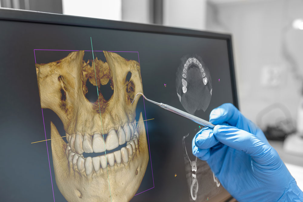
A standard dental X-ray can show a great deal, but some tooth problems hide behind overlapping roots, dense bone, or complex internal anatomy. In selected cases, CBCT imaging for root canal treatment gives an endodontist a three-dimensional view that can make diagnosis and planning more precise. Lake Houston Endodontics uses advanced dental technologies in Humble, TX to investigate difficult symptoms and plan specialized care.
A CBCT scan is not needed for every toothache or every root canal. Instead, it is used when the additional information may change the diagnosis, reveal anatomy that cannot be seen clearly on two-dimensional images, or help the endodontist choose between treatment options. Understanding what the scan does can help patients feel more confident when CBCT imaging for root canal treatment is recommended.
What Is a Dental CBCT Scan?
CBCT stands for cone beam computed tomography. The scanner moves around the patient’s head and captures many small images from different angles. Computer software then combines those images into a three-dimensional view of the teeth, roots, jawbone, and nearby structures.
The resulting data can be viewed in thin slices from several directions. An endodontist may rotate the image, enlarge a particular root, or examine the area from the side, front, and top. This flexibility is valuable because root canal systems are three-dimensional, even though traditional dental X-rays flatten the area into a two-dimensional picture.
CBCT imaging for root canal treatment is focused on diagnosis and planning. It does not replace a clinical examination, pulp testing, bite testing, or standard digital X-rays. Instead, it adds another layer of information when the basic findings leave an important question unanswered.
How CBCT Differs From a Standard Dental X-Ray
A standard dental X-ray is quick, useful, and appropriate for many endodontic evaluations. It can show decay, large restorations, bone changes, previous root canal fillings, and the general shape of roots. However, structures can overlap. A root positioned behind another root may be difficult to separate, and a small area of bone loss may not be visible from the angle used.
CBCT imaging for root canal treatment reduces the problem of overlap by displaying the area in three dimensions. For example, the endodontist may be able to see whether a dark area is associated with one root or another. The scan may also show that a root curves in a direction that was hidden on the standard image.
Still, more imaging is not always better. A standard X-ray may already provide everything needed for a straightforward case. The endodontist chooses CBCT when the expected diagnostic value justifies the scan.
Finding Extra or Unusual Root Canals
Teeth do not all follow a simple blueprint. A molar may have an extra canal, a canal may divide and reconnect, or roots may curve sharply. These variations can be difficult to identify on a flat image. If infected tissue remains in an untreated canal, symptoms or disease may continue after treatment.
CBCT imaging for root canal treatment can help map unusual anatomy before the procedure begins. This gives the endodontist a clearer idea of where to look and how the canals may travel through the root. Magnification and illumination are then used during treatment to locate and clean the actual openings.
The scan does not perform the treatment, and it cannot guarantee that every microscopic branch will be visible. However, a three-dimensional roadmap can make a complex case more understandable and support a more efficient plan.
Evaluating a Tooth That Still Hurts After a Root Canal
A previously treated tooth may become painful months or years later. Causes can include a missed canal, leakage from a restoration, new decay, a fracture, or persistent inflammation around a root. Because root filling materials and crowns can make standard images harder to interpret, additional views may be useful.
CBCT imaging for root canal treatment can show the location and size of bone changes around individual roots. It may also reveal untreated anatomy or a problem that affects only one side of a tooth. These details help the endodontist determine whether endodontic retreatment, surgery, monitoring, or another option deserves consideration.
Not every source of pain appears on a scan. Bite problems, muscle pain, early cracks, and some inflammatory conditions may require clinical testing and careful history. Therefore, imaging is interpreted as part of the whole examination rather than treated as the only answer.
Planning Endodontic Surgery
When inflammation remains near a root tip after nonsurgical treatment, an apicoectomy or another form of endodontic surgery may be considered. Before surgery, the endodontist needs to understand the root’s position, nearby bone, and relationship to surrounding anatomy.
CBCT imaging for root canal treatment and surgery can help identify which root is involved and where the problem is located. It may also show the thickness of bone over the root or the proximity of nearby structures. This information supports a more targeted surgical approach.
Patients can learn more about endodontic surgery in Humble, TX when nonsurgical treatment alone cannot address the problem. The recommendation depends on the tooth, restorability, and expected long-term outlook.
Can CBCT Find a Cracked Tooth?
Cracks are among the most challenging dental problems to diagnose. A large fracture or the bone pattern associated with a root fracture may be visible on CBCT. However, very fine cracks can still be too small to appear clearly. Restorations and root filling materials may also create visual artifacts that make interpretation harder.
For this reason, CBCT imaging for root canal treatment is only one part of a cracked tooth evaluation. Bite testing, magnification, transillumination, gum measurements, and symptom patterns may provide important clues. Pain when releasing a bite, isolated deep gum probing, or a recurring gum bump can influence the diagnosis even if the crack itself is not visible.
A scan can still be valuable because it may rule in or rule out other causes. For example, it may show a missed canal or localized bone loss that changes the treatment discussion.
Benefits of CBCT Imaging for Root Canal Treatment
- Three-dimensional detail: The scan allows roots and surrounding bone to be viewed from multiple directions.
- Improved anatomy mapping: CBCT imaging for root canal treatment may reveal extra canals, unusual curves, or root variations.
- More focused retreatment planning: The endodontist can identify which root or region may be associated with persistent disease.
- Better surgical preparation: A scan can clarify the location of a root tip and nearby structures before endodontic surgery.
- Support for difficult diagnosis: CBCT may provide useful information when symptoms continue but standard images are inconclusive.
- Clearer patient communication: Three-dimensional views can help patients understand why a treatment option is being recommended.
What the Scan Experience Is Like
CBCT imaging is generally quick. You may stand or sit while the machine is positioned around your head. The team asks you to remain still, and the scanner rotates around the area. Nothing is placed deep inside the mouth, although positioning supports may be used to help you stay steady.
The scan itself does not hurt. After the image is captured, the endodontist reviews the data on a computer. In many cases, the most relevant views can be shown during the consultation so you can see the roots and surrounding bone.
Metal jewelry, removable appliances, or other objects near the head may interfere with the image. Follow the team’s instructions about removing earrings, glasses, or removable dental items before the scan.
How Imaging Is Selected Thoughtfully
CBCT imaging for root canal treatment is ordered when the expected benefit is meaningful. The field of view can often be limited to the region being evaluated, rather than scanning a larger area than necessary. The dental team also considers whether previous imaging is available and whether a standard X-ray can answer the question.
Patients should tell the office if they are pregnant or may be pregnant before imaging. They should also mention recent scans so the endodontist can determine whether those records may be useful. If another dental office has already taken a CBCT scan, ask whether the file can be transferred for review.
It is reasonable to ask why the scan is recommended. A clear answer might include locating an extra canal, evaluating a root fracture, measuring a bone lesion, or planning surgery. The reason should be connected to a specific diagnostic or treatment decision.
How CBCT Works With Microscopes and Digital X-Rays
Endodontic technology is most effective when tools complement one another. Digital X-rays provide efficient two-dimensional views. CBCT offers three-dimensional context. Meanwhile, a dental microscope gives high magnification and illumination inside the tooth during treatment.
For example, CBCT imaging for root canal treatment may suggest that a narrow extra canal is present. The microscope then helps the endodontist search for the canal opening. Ultrasonic instruments may be used to remove small amounts of calcified material while preserving surrounding tooth structure.
No technology replaces training and judgment. The endodontist must decide which finding matters, whether it matches the symptoms, and whether treatment can improve the tooth’s outlook.
Frequently Asked Questions About CBCT Imaging for Root Canal Treatment
Does every root canal need a CBCT scan?
No. Many root canals can be diagnosed and treated with an examination and standard digital X-rays. CBCT is used when three-dimensional information may improve diagnosis or planning.
Can a CBCT scan show infection?
It can show changes in the bone that may be associated with inflammation or infection. The endodontist combines those findings with symptoms, testing, and the condition of the tooth.
Will CBCT always show a crack?
No. Some cracks are too fine to be seen directly, and metal or filling materials can affect the image. Clinical tests remain important in a cracked tooth evaluation.
Is the scan uncomfortable?
The scan is generally quick and painless. The main requirement is remaining still while the machine rotates around the head.
Can an old CBCT scan be used?
Possibly. The value depends on when it was taken, the area included, image quality, and whether the tooth has changed since then. The endodontist can review the file and decide whether new imaging is needed.
See the Full Picture Before Treatment
CBCT imaging for root canal treatment can reveal details that are hidden by overlapping anatomy on a standard X-ray. When used selectively, it helps an endodontist investigate complex pain, plan retreatment, evaluate surgery, and explain findings more clearly. The scan is most useful when it answers a specific question and is interpreted alongside a complete clinical examination.
For advanced endodontic diagnosis in Humble, TX, contact Lake Houston Endodontics. Call Us Today at (832) 777-6056 or contact the practice online to schedule an appointment and learn whether CBCT imaging for root canal treatment may be appropriate for your tooth.